Wondering how often you should have a dental check-up? Although the answer is usually „once a year”, the key is not only how often you visit the surgery, but more importantly - what happens during that visit.
A regular and, most importantly, thorough dental check-up is the absolute basis of prevention and the most effective way to avoid pain and costly treatment. A comprehensive dental examination is much more than a quick look inside the mouth.
In this article, we'll explain the key components of a check-up visit that make a real difference to your health. We will also show you why it is the best investment in a beautiful and, most importantly, healthy smile for years to come.
What does an in-office dental review include?
The first and fundamental step of any professional dental consultation is a detailed clinical examination. This is the moment when the dentist assesses all the visible structures of the oral cavity. During this part of the visit, the dentist carefully checks several key areas to get an initial picture.
What is assessed during a clinical trial?
- Tooth condition: The doctor carefully views each tooth for foci of caries, The patient's teeth should be examined for signs of wear, fractures in the enamel or excessive abrasion. It is equally important to assess the tightness and condition of existing fillings, crowns or bridges, as these may need to be replaced over time.
- Gum and periodontal health: Redness, swelling or bleeding of the gums during the examination are warning signs that may indicate inflammation or the beginnings of periodontitis (a disease that leads to tooth loss).
- Presence of scale and sediment: Dental deposits harbour bacteria, so their location and quantity are important diagnostic information.
- Mucosal condition: Inspection of the tongue, the inside of the cheeks and the palate allows early detection of worrying changes that may be relevant to overall health.
This is a key stage that allows an initial check of the condition of the teeth, but to get the full picture we need to look deeper - where the human eye cannot reach.
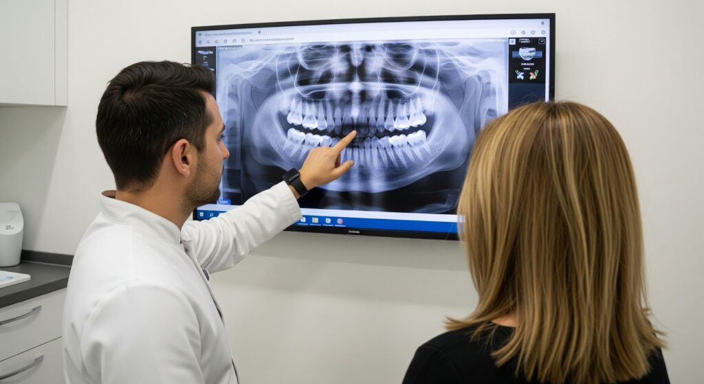
X-ray diagnosis - what can be seen on a pantomographic image?
Many of the most serious dental problems develop painlessly and in secret. Tooth decay, inflammation around the roots or retained eights produce no symptoms for a long time. Therefore, modern dental diagnostics cannot exist without X-ray examinations.
The key and fundamental imaging examination is the pantomogram, or panoramic radiograph of the teeth. This single X-ray shows all the teeth, the jaw, the mandible and the temporomandibular joints. As confirmed by specialists, it is an examination of great diagnostic value, allowing the evaluation of structures that are not visible during intraoral examination.
What can a pantomogram detect?
- Caries previously untreated teeth and under leaking fillings.
- Retained teeth (usually figure-eights) and their position in relation to neighbouring structures.
- Inflammatory lesions at the roots of teeth, such as cysts or granulomas, which can cause pain or complications in the future.
- Level and condition of alveolar bone, which is crucial in the diagnosis and monitoring of periodontitis.
- Root abnormalities and possible changes in the temporomandibular joints.
Sometimes, the changes detected on the pantomogram form the basis for a deeper diagnosis of the detected abnormalities and detailed imaging of the detected abnormalities, such as wing-bite radiographs or cone tomography.
Full picture of the state of the oral cavity
It is only by combining the in-depth examination in the office with the information provided by the pantomogram that the doctor is able to gain a complete and reliable picture of the patient's oral health. This synergy not only allows you to accurately plan your eventual treatment, but above all gives you confidence that nothing has been overlooked.
Let us also remember that oral health is inextricably linked to the health of the whole body. Therefore, regular and thorough check-ups are an investment in your overall well-being.
Frequently Asked Questions (FAQ)
1. Is the pantomographic examination safe?
Yes. Today's digital cameras use a minimal, precisely targeted radiation dose that is many times lower than in older generation cameras. It is a completely safe examination and the diagnostic benefits far outweigh any risks.
2 How often should a pantomogram be performed?
The decision is always made by the dentist based on the individual needs of the patient. In patients without specific problems, as a review examination, it is usually performed every 2-3 years. In patients undergoing orthodontic or implant treatment or with suspected multiple problems, more frequent radiographs may be necessary.
3. How do I prepare for a dental review?
The visit does not require any special preparations. It is sufficient to brush your teeth thoroughly before the appointment. It is also a good idea to remind yourself of the history of any previous treatments and a list of medications you are taking.

Benefit from a comprehensive diagnosis
Make an appointment for a dental check-up at the Modern Dental & Orthodontics Clinic in Warsaw's Wola district today.