
Modern dentistry is built on precision. For treatment to be effective, safe and comfortable, the clinician must look deeper — where the eye alone cannot reach.
At our clinic we combine experience with modern technology, and we know how crucial an accurate diagnosis is.
In this article we will guide you through the most important types of radiological examination so that you know what to expect and why they are essential.
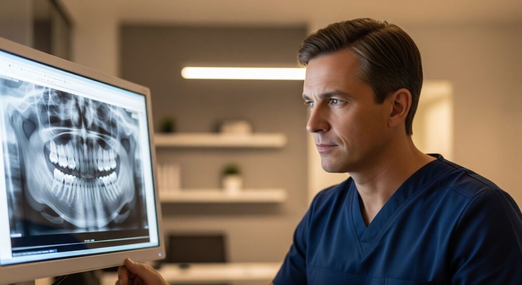
Periapical (point) radiograph — precision on a small scale
The periapical radiograph, also called a point radiograph, is the most frequently taken image in the dental clinic. It is small but extremely detailed — it shows one to three teeth and the bone surrounding them.
When is it essential? It is the first-choice investigation for a suspected specific problem. We take it to:
- Confirm and assess the depth of a carious lesion, especially on the proximal (contact) surfaces of the teeth.
- Check the condition of a restoration (filling).
- Conduct root canal treatment precisely — a periapical radiograph is required before, during and after the procedure to assess the length and accuracy of canal preparation.
- Detect inflammatory changes around the root apex, such as cysts or granulomas.
Its greatest advantage is high image resolution at a very low radiation dose. It is a quick and effective method for diagnosing a specific tooth problem.
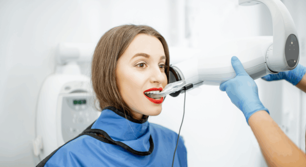
Bitewing radiograph — a guardian in the fight against caries
This is a specialist dental radiograph that images the crowns of the upper and lower teeth simultaneously in a single exposure. The patient bites on a small disposable holder that holds the film or sensor in position.
This examination is invaluable in prevention. It allows the very early detection of cavities — before they become extensive and begin to cause pain.
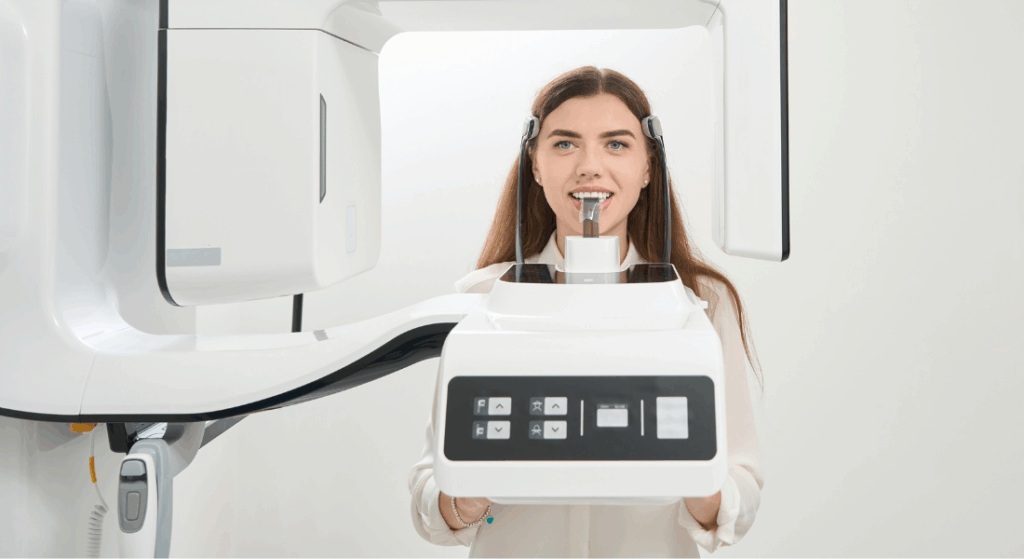
Panoramic radiograph (OPG) — a broad view of your smile
A panoramic radiograph is a wide-field dental X-ray that gives an overall picture of the entire oral cavity. In a single image one can see all the teeth (including impacted teeth and developing tooth buds), the mandible, the maxilla, the maxillary sinuses and the temporomandibular joints.
When do we take a panoramic radiograph?
- During the first visit, to obtain a full picture of the dental condition and plan comprehensive treatment.
- Before starting orthodontic treatment, to assess the position of all teeth and their developing buds.
- In planning surgical procedures, especially wisdom tooth extraction.
- In implant and prosthetic diagnostics, to evaluate bone conditions.
- When periodontal disease or pathological changes in the bone are suspected.
At our clinic the panoramic radiograph is a fundamental diagnostic tool that enables us to create precise and effective treatment plans.
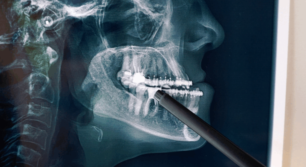
Cephalometry (lateral skull radiograph) — the cornerstone of orthodontics
Cephalometry is a specialist radiographic image that shows the lateral profile of the skull. It reveals the spatial relationship between the maxillary and mandibular bones, the inclination of the teeth and the proportions of the face.
This examination is absolutely key in modern orthodontics. Thanks to precise measurements made from the cephalogram, the orthodontist can plan the direction and extent of tooth and jaw movements.
Cephalometry is also taken before planned orthognathic surgery. If you are considering fixed braces, the cephalogram will be one of the first investigations requested.
Cone-beam computed tomography (CBCT) — three-dimensional precision
Cone-beam computed tomography (CBCT) is the most advanced imaging examination in dentistry. Unlike flat 2D images, CBCT generates a three-dimensional model that can be rotated and analysed from every angle.
CBCT is essential in:
- Implantology: for precise planning of implant placement, assessment of bone thickness and density, and avoidance of damage to critical structures (the inferior alveolar nerve, the maxillary sinus).
- Endodontics: in complex root canal treatment cases, for locating additional or curved root canals.
- Dental surgery: in planning the removal of impacted teeth, especially when they lie close to the inferior alveolar nerve.
- Diagnosis of temporomandibular joint disorders and pathological lesions.
Thanks to our in-house CS 8200 3D scanner, we can offer the full range of radiological services on site. This ensures fast diagnostics and the ability to create a treatment plan during a single visit.
Modern diagnostics for your safety and comfort
From the precise periapical radiograph, through the panoramic image, to advanced 3D tomography — every type of radiological examination has its unique role in looking after your oral health.
At the Klinika Modern Dental & Orthodontics in Wola, Warsaw, we take a comprehensive approach. Having state-of-the-art diagnostic equipment in-house means that we do not have to refer you elsewhere for imaging — everything is done in one place, on one visit.
Take the first step on your journey to a beautiful smile. Book a consultation appointment and we will take care of the rest. We look forward to hearing from you!
Frequently Asked Questions (FAQ)
1. Are dental radiographs safe?
Modern digital X-ray equipment emits a minimal, precisely targeted radiation dose that is safe for the health of adult patients. As an additional safeguard, a protective lead apron is always used during the examination.
2. How often can dental radiographs be taken?
The frequency of radiographic examinations is always dictated by diagnostic needs and prescribed by the clinician. There are no rigid rules, but as a standard, a panoramic radiograph is recommended once every 1–2 years.
3. Do I need to prepare in any special way for a dental radiograph?
Before the examination you will be asked to remove metal objects from the head and neck area (glasses, earrings, necklaces, removable dentures). No other preparation is required.
4. Can a pregnant woman have a dental radiograph?
As a rule, radiological examinations are avoided in pregnant women, especially in the first trimester. In situations of absolute necessity, it is possible — but only with the use of a protective lead apron on the abdomen and only on the clinician's recommendation.