
What sets a fractured tooth root apart from other causes of pain after root canal treatment
- A fractured tooth root, known clinically as a vertical root fracture (VRF), is a crack running along the root as far as the canal. It is a diagnosis in its own right, not a variety of cracked crown. The other four types of crack are covered in a separate article.
- It usually declares itself years after root canal treatment, and for a long time it causes no symptoms at all.
- Three signs narrow the suspicion: a narrow, deep periodontal pocket at one surface of the tooth, a sinus tract close to the gum margin, and pain on biting despite correctly performed treatment.
- Cone beam computed tomography (CBCT) does not show the crack itself. It shows the shape of the bone loss around the root, and it is that shape which guides the diagnosis.
- Once dentine has cracked it does not heal back together, and no strong studies confirm any method of repair. In a single-rooted tooth what remains is extraction; in a multi-rooted tooth, sometimes amputation of the fractured root alone.
Pain after root canal treatment most often does not mean a fracture; behind it there is usually something that can be put right: a filling left too high, a canal that was not found at the first attempt, or inflammation at the root tip that has not yet settled. Within that group, vertical root fracture is the less common diagnosis and at the same time the one that cannot be reversed.
The name “cracked tooth” covers at least five different diagnoses: craze lines in the enamel, a fractured cusp, a crack in the crown reaching the dentine, a split tooth, and vertical root fracture. This article concerns only the last of them, that is a cracked tooth after root canal treatment. The question at issue here is the one asked frequently once that diagnosis is made: if the tooth has already had root canal treatment, why will further treatment change nothing.
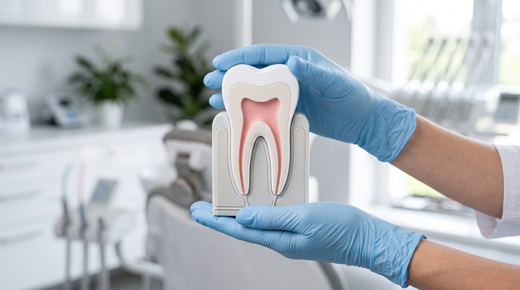
Why is a fractured tooth root a different diagnosis from a cracked crown?
The difference is not made by depth alone, though depth differs too. It is made by the direction of the crack and by whether it can be sealed.
A crack in the crown starts on the biting surface and travels down into the dentine; as long as it does not pass below bone level, it stays within the area the dentist can see, can clean and can rebuild. In its 2025 position statement the European Society of Endodontology reports encouraging outcomes for such teeth, on condition that they receive a restoration covering the cusps, that occlusal overload is brought under control, and that there is no deep pocket of periodontal origin alongside them.
A vertical root fracture runs differently. It follows the long axis of the root and involves the cementum covering the root, the dentine and the canal lumen all at once. It usually begins at the cervical part of the root or at the root tip, and for a long time nothing of it can be seen from outside. The same position statement lists it among the diagnoses with a poor prognosis, for which it recommends extraction, while in a multi-rooted tooth it allows removal of the affected root alone.
The difference has one more layer, and that one matters more for the decision. A cracked crown is a problem of seal: infection enters the pulp through the crack, and that infection can be removed. A fractured tooth root is a problem of structure. The crack becomes a route by which bacteria descend into the periodontium, and at the same time the place where the root goes on splitting under the pressure of the bite. Once started, the crack does not stop of its own accord. The reason lies in the material itself: a crack forms readily in dentine, and what ought to arrest its progress proves too weak under loading repeated with every mouthful. That is how a biomechanical review in the Journal of Dentistry describes it.
Three signs that narrow the suspicion
None of them is enough on its own. Together, though, they change the question from “why does this tooth hurt” to “could this be a fracture”.
A narrow, deep pocket at one surface of the tooth
This is the strongest of the signs. A deep periodontal pocket raises the odds of a vertical root fracture more than tenfold. With no other finding is the difference that large. The meta-analysis covered 2877 teeth, of which 489 had a confirmed fracture. Ratios of this kind say how much the suspicion rises, not how certain it is. And the estimate itself is wide. An isolated pocket calls for the tooth to be examined with this in mind; on its own it does not settle the diagnosis. What decides is not depth alone but shape: periodontal disease produces a broad defect spread around the tooth, whereas a fracture leaves a gap as narrow as the probe itself, at one surface, with the neighbouring tooth entirely healthy. A narrow defect was present in 78 per cent of the fractured teeth in a Japanese series of 288 teeth with an isolated pocket of at least five millimetres. A pocket detectable on both sides of the same root is by then a decisive finding.
An isolated deep pocket at a single tooth after root canal treatment is not an ordinary periodontal problem, and subgingival debridement on its own will not close it. Before anyone begins to treat it, the dentist probes the whole circumference of the tooth.
A sinus tract close to the gum margin
A sinus tract at a tooth is a small channel through which pus finds an outlet on the gum. With a fracture it appears, across different series, in 18 to 67 per cent of teeth, and it is recognised by where it lies: not at the root tip, as in an ordinary periapical infection, but higher up, at mid-root level or right at the gum margin. Two sinus tracts at one tooth argue for a fracture particularly strongly. The presence of a sinus tract on its own is associated with close to five times higher odds of a vertical root fracture - that is what the same meta-analysis found.
Pain on biting despite correctly performed treatment
The weakest of the three signs, and the one most often misread. Tenderness of the tooth to tapping accompanies a fracture more often than it accompanies its absence, but the difference is small: the odds rose less than twofold. That figure does not apply to pain on biting itself. Its value lies elsewhere. If a tooth hurts despite root canal treatment that was performed correctly, and one of the two preceding signs joins it, the suspicion of a fracture rises. The meta-analysis, however, counted each finding separately and does not give the strength of such a combination.
Who is at risk?
The risk concerns above all teeth that have had root canal treatment, and in them a fracture usually declares itself years after the procedure.
Vertical root fractures do also occur in teeth that have never had root canal treatment, but rarely. Most diagnoses concern root filled teeth, and the frequency reported for that group falls between 4 and 32 per cent. This does not mean that every root filled tooth will fracture one day - in the great majority nothing of the kind happens. The spread probably comes from fracture having been diagnosed differently in different papers; that is how a 2022 review accounts for it. The figures also depend on where they were collected: it comes out one way in general practice and another in an endodontic clinic. Among teeth referred for surgery, a vertical root fracture is confirmed in just under one third. That was counted on 411 teeth in a Thai centre.
Three features recur across successive papers, although not all of them were confirmed by the meta-analysis:
- age: fractures become more frequent after the age of forty;
- the type of tooth: molars and premolars, that is the teeth taking the greatest load;
- how much dentine is left in the root: in the Thai study the odds rose by half when the lumen of the prepared canal exceeded one third of the width of the root.
Only the last of the three depends on how the treatment was carried out, and it is the one that explains today's emphasis on conserving dentine.
It is worth saying plainly what is not known. For years it was repeated that canal preparation itself creates microcracks in the dentine, and that everything starts from those. The conclusion came from studies in which the root was cut into slices. Micro-computed tomographic analyses and work on cadaver material do not confirm it: the microcracks were either there beforehand, or arose when the tooth was extracted and then dried out. The difference is not academic. If microcracks really were born under the instrument, canal preparation itself would be to blame; that has not been shown. What remains is the factor that could be counted: how much dentine is left in the root.
How is the diagnosis confirmed?
Not by a single examination. By a route that has four stages and gives certainty only at the last of them.
- Suspicion: a clinical sign, or an incidental finding on a follow-up radiograph.
- The dentist walks the probe around the whole circumference of the tooth in small steps. Without this step an isolated pocket is easily missed, because it is met at one spot only.
- A radiograph, and where the result is equivocal, cone beam computed tomography. Both examinations serve to assess the shape of the bone loss around the root, not to see the crack itself.
- The dentist inspects the root directly: under the microscope, after the restoration has been removed, and sometimes only once the root has been exposed surgically or the tooth has already been taken out.
CBCT is what disappoints most often. It detects roughly four fractures in five, and roughly one tooth in five without a fracture gets a false signal from it. The pooled results, measured against direct inspection of the root, gave a sensitivity of 0.78 and a specificity of 0.80. The reason is physical: the crack is usually between 50 and 100 micrometres wide, that is below the resolution of CBCT, and gutta-percha and a metal post distort the image further.
A normal CBCT image therefore does not rule out a fracture.
What CBCT does show well is something else: the shape of the bone defect around the root, and with a fracture that shape can be characteristic, looking like a halo in the form of the letter J. An isolated radiolucency alongside the lateral wall of the root means four times higher odds of a fracture - those data again come from the same Thai series. It is therefore worth asking for an endodontic consultation even when the previous radiograph showed nothing.
In everyday clinical practice at Modern Dental & Orthodontics (Klinika MDO) we meet the question of why a dentist holding a finished CBCT report still does not say “yes” or “no”. Because an examination of that kind shifts the probability up or down, and does not give certainty.
Why a fractured root cannot today be bonded back together or treated out
A fractured root cannot today be bonded back together in any way of proven effectiveness: dentine does not heal back together, and the crack becomes colonised by bacteria.
A fractured bone heals because it is living tissue and rebuilds on both sides of the break. Root dentine cannot do that. A second problem is added to it: from the moment the crack has connected with the mouth, it is filled by biofilm, an organised layer of bacteria that cannot be removed from a space the width of a hair. Root canal retreatment and root-end surgery make the same mistake: they remove the infection from the canal, and the crack remains.
In practice the order is sometimes the reverse of the textbook one. Certainty about a fracture is sometimes available only once root canal retreatment has already been carried out and has not helped. That need not mean anyone made a mistake: no imaging examination gives certainty before the tooth is opened. What counts is the moment at which the fracture stops being a suspicion. From that moment a further endodontic procedure no longer has any justification.
Two main approaches have been tried to save such teeth: dentists either took the tooth out, bonded the fragments together with resin outside the mouth and replanted it, or reached the crack through the gum and filled it with cement. The results of these attempts cannot be pooled today: eight publications describing six studies are all case series of differing methodology, most of them without a comparison group. The authors of a 2025 review close cautiously and unambiguously - the evidence is sparse and of low quality, so nothing certain can be stated about the effectiveness of these techniques. For a patient it comes to this: nobody today can give the chance that a tooth bonded in this way will survive.
Neither a further root canal treatment nor root-end surgery is performed “on a trial basis” on a tooth in which a fracture has been diagnosed. Such a procedure removes further dentine and puts the decision off, and it does not improve the prognosis.
What happens once the diagnosis is confirmed?
Most often the tooth has to be extracted. In a tooth with a single root, a fractured tooth root leaves no other predictable route, while in a multi-rooted tooth there is an intermediate option.
Molars sometimes offer a way out in between: the fractured root alone can be removed and the others kept. The procedure is called root amputation, and in the variant where the tooth is divided into two parts - hemisection. The conditions are that the fracture does not reach the furcation, that the roots which remain have healthy periodontal tissue, and that enough tooth is left after the amputation for a restoration to be seated on it. It has to be added, though, how much is known about this: nobody has counted how long teeth serve after a root has been amputated for a fracture specifically. The figures quoted for the method, more than nine teeth in ten at ten years, come from root amputations carried out for periodontal reasons, in teeth weakened in a different way.
The appointment is not put off, and the reason is a concrete one. For as long as the crack remains in the mouth, the bone defect along the root grows, and it is that bone which later has to hold the restoration. The position statement of the European Society of Endodontology says it plainly: delay after diagnosis increases the risk of an acute flare-up and of further bone loss, which may complicate or delay implant treatment. That is why the restorative plan is discussed before the extraction.
Nor is this a rare ending. Among 1564 root filled teeth that were replaced by implants, vertical root fracture accounted for about one fifth of the extractions and was the third reason, after secondary caries and crown fracture. A separate article sets out whether to save the tooth or place an implant.
Frequently asked questions
Does a fractured tooth root always hurt?
No. Early on, a fracture usually causes no symptoms at all and is sometimes picked up incidentally on a follow-up radiograph. Pain on biting appears later and is the weakest of the signs; the related tenderness to tapping raised the odds of the diagnosis less than twofold in the meta-analysis. Absence of pain rules nothing out, so follow-up radiographs after root canal treatment matter.
My tooth hurts despite root canal treatment. Is that always a fractured tooth root?
Most often not. Pain after root canal treatment usually has other causes: a filling left too high, a canal missed at the first attempt, inflammation at the root tip that has not settled, or a crack in the crown itself. Suspicion of a root fracture rises only once an isolated deep pocket or a sinus tract at the gum margin joins the pain.
Can the fracture be seen on an ordinary radiograph?
Rarely. A radiograph is a flat image, and the crack is so narrow that it shows only when the beam falls exactly along it. What can be seen are the consequences: widening of the periodontal ligament space along the root, or a radiolucency in the form of the letter J. Their absence settles nothing, and their presence raises the suspicion without confirming it.
Does this mean the root canal treatment was done badly?
Usually not. Two of the three factors - the patient's age and the type of tooth - are within nobody's control. The only one the treatment influences is the amount of dentine removed from the root, and that is why canals are prepared more sparingly today than they used to be. Correctly performed root canal treatment is not in itself regarded as a cause of fracture, although root filled teeth fracture more often than vital ones.
Does a post and core increase the risk of fracture?
The papers do not agree. A 2023 meta-analysis showed no association between the post itself and fracture, while a more recent clinical study pointed not to the post but to the width of the post space drilled for it: the odds rose when it exceeded one third of the width of the root. What matters, then, is the amount of dentine removed rather than the presence of a post and core as such.
How quickly does such a tooth have to be extracted?
Without delay, from the moment the diagnosis is certain, although this is not an emergency. Every week with the crack in the mouth means further loss of bone around the root. What does require immediate care is increasing swelling of the face, fever, or difficulty swallowing or opening the mouth.
Can an implant be placed straight away after the extraction?
That is sometimes possible, though what decides it is the state of the bone after the extraction, not the plan made beforehand. With a fracture the buccal bone plate is sometimes already destroyed, which can be seen on CBCT before the procedure. The final assessment is made by the dentist planning the restoration, after inspecting the empty socket.
When to suspect a fracture: treatment from years ago, pain in one spot, a gum with a history
Three questions weigh most heavily: when the tooth had root canal treatment, whether the complaint always comes back in the same place, and whether the gum beside it has ever swollen or discharged. With a vertical root fracture the answers fall into a recurring pattern: treatment from some years back, pain returning at one point, a gum with a history.
An isolated deep pocket at a single tooth after root canal treatment calls for the whole circumference to be checked with a probe, before anyone begins to treat it as a periodontal problem. Increasing swelling of the face with fever, or with difficulty swallowing, is an indication for an urgent appointment the same day, whatever the earlier diagnosis.
Read more:
- Tooth Extractions in Warsaw Wola
- A cracked tooth — when it can be saved and when it must be removed
- Root canal retreatment after failed treatment — when it makes sense and when it does not
- Save the tooth or place an implant — how to make a decision you will not regret
Content and liability disclaimer
This article is informational and educational in nature and does not constitute medical advice, a diagnosis or a therapeutic recommendation — it does not replace a consultation with a specialist. If you are experiencing symptoms, have doubts or are facing a decision about treatment, consult a dentist. The methods described and the data cited are general in nature. The results of scientific studies relate to populations, not to an individual patient. The course and outcome of therapy depend on the individual clinical situation and may vary between patients. No information contained in this article constitutes a guarantee of result. The content has been prepared with due care, based on publicly available medical knowledge and the scientific publications indicated in the Sources section. We do not advise taking or refraining from any health-related action solely on the basis of the content of this article, without prior consultation with a doctor.
Sources
Source 1
Links https://doi.org/10.1111/iej.14186 │ https://pubmed.ncbi.nlm.nih.gov/39840523/
Description Patel S, Teng PH, Liao WC, Davis MC, Fidler A, Haupt F, Fabiani C, Ordinola Zapata R, Bose R. „Position statement on longitudinal cracks and fractures of teeth.” International Endodontic Journal. 2025;58(3):379-390.
Source 2
Links https://doi.org/10.1111/iej.13737 │ https://pubmed.ncbi.nlm.nih.gov/35338655/
Description Patel S, Bhuva B, Bose R. „Present status and future directions: vertical root fractures in root filled teeth.” International Endodontic Journal. 2022;55(Suppl 3):804-826.
Source 3
Links https://doi.org/10.1016/j.joen.2023.06.004 │ https://pubmed.ncbi.nlm.nih.gov/37307871/
Description Haupt F, Wiegand A, Kanzow P. „Risk Factors for and Clinical Presentations Indicative of Vertical Root Fracture in Endodontically Treated Teeth: A Systematic Review and Meta-analysis.” Journal of Endodontics. 2023;49(8):940-952.
Source 4
Links https://doi.org/10.1016/j.joen.2025.07.020 │ https://pubmed.ncbi.nlm.nih.gov/40769327/
Description Promchouy P, Chotvorrarak K, Osiri S. „Prevalence of Vertical Root Fractures in Root Filled Teeth and Their Clinical and Radiographic Risk Factors: A Modern Case-Control Analysis Based on Surgical Diagnosis.” Journal of Endodontics. 2026;52(1):68-78.
Source 5
Links https://doi.org/10.1016/j.joen.2021.04.022 │ https://pubmed.ncbi.nlm.nih.gov/33984375/
Description PradeepKumar AR, Shemesh H, Nivedhitha MS, Hashir MMJ, Arockiam S, Uma Maheswari TN, Natanasabapathy V. „Diagnosis of Vertical Root Fractures by Cone-beam Computed Tomography in Root-filled Teeth with Confirmation by Direct Visualization: A Systematic Review and Meta-Analysis.” Journal of Endodontics. 2021;47(8):1198-1214.
Source 6
Links https://doi.org/10.1016/j.joen.2023.08.018 │ https://pubmed.ncbi.nlm.nih.gov/37660764/
Description Lee K, Ahlowalia M, Perez Alfayate R, Patel S, Foschi F. „Prevalence of and Factors Associated With Vertical Root Fracture in a Japanese Population: An Observational Study on Teeth With Isolated Periodontal Probing Depth.” Journal of Endodontics. 2023;49(12):1617-1624.
Source 7
Links https://doi.org/10.1016/j.joen.2023.12.002 │ https://pubmed.ncbi.nlm.nih.gov/38141831/
Description Kim DH, Tawil PZ, Albouy JP, Duqum I. „Retrospective Assessment of Endodontically Treated Teeth Replaced by Dental Implants.” Journal of Endodontics. 2024;50(3):310-315.
Source 8
Links https://doi.org/10.1016/j.joen.2025.05.004 │ https://pubmed.ncbi.nlm.nih.gov/40374035/
Description Ran S, Wang Q, Wang J, Huang J, Zhou W, Zhang P, Yuan K, Cheng Y, Gu S, Zhu J, Huang Z. „Diagnosis of In Vivo Vertical Root Fracture in Endodontically Treated Teeth Using Machine Learning Techniques.” Journal of Endodontics. 2025;51(10):1393-1404.
Source 9
Links https://doi.org/10.1016/j.jdent.2023.104424 │ https://pubmed.ncbi.nlm.nih.gov/36657703/
Description Chen S, Arola D, Ricucci D, Bergeron BE, Branton JA, Gu LS, Tay FR. „Biomechanical perspectives on dentine cracks and fractures: Implications in their clinical management.” Journal of Dentistry. 2023;130:104424.
Source 10
Links https://doi.org/10.1111/aej.12923 │ https://pubmed.ncbi.nlm.nih.gov/39891472/
Description Corbella S, Taschieri S, Tsesis I, Goldberger T, Francetti L, Rosen E. „The Efficacy of Surgical Techniques for the Management of Confirmed Vertical Root Fractures: A Systematic Review.” Australian Endodontic Journal. 2025;51(1):218-229.