
It does not hurt because it has a stoma — and that is bad news
- A lump on the gum that alternately discharges and scars over requires an appointment this week.
- A dental sinus tract stops hurting because it has an outlet: the stoma works like a valve that lets the pus out and takes the pressure off.
- The stoma closes on its own and comes back on its own, and the lump disappearing settles nothing. Rinsing, ointment or an antibiotic alone do not close the matter.
- Most often the stoma does not lie over the diseased tooth but next to it, and which tooth is discharging is settled by a radiograph taken with a gutta-percha cone in the tract.
- This article concerns a chronic condition with a patent stoma. A swelling cheek and fever are already a tooth abscess, which is a different level of urgency.
Where does a dental sinus tract come from?
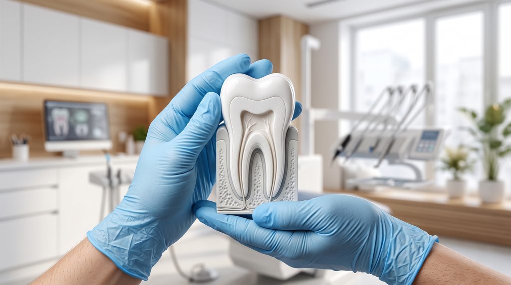
From an infection in the root canal of one of the teeth in the area. The pulp, the living tissue that fills the inside of the tooth, dies after deep decay or after an injury, and the canal left without it has no blood supply, so neither immune cells nor a drug taken by mouth reach it. Infection also remains after root canal treatment that did not remove it completely.
The infection travels down through the opening at the root apex into the bone and an inflammatory focus forms there, called a periapical lesion in radiographic reports; bone around the apex is lost and the pus looks for a way out. It finds one where the resistance is lowest: it breaks through the bone and opens onto the surface of the gum. That drainage channel is the dental sinus tract, and the visible opening with a small collar of tissue around it is called the stoma. A sinus tract on the gum, a hole in the gum and a pimple that will not heal are everyday names for the same stoma.
The sinus tract is not a permanent structure: most often it has no lining of its own and closes by itself as soon as the reason it formed disappears. That is good news and at the same time the source of the commonest mistake, because if the stoma closes on its own after treatment, it also closes on its own without treatment - and from the outside it looks identical. A dental sinus tract is therefore a symptom, not a disease: it is the route by which the infection comes out.
Even periapical lesions that look self-sustaining contain bacteria, and the route to healing is root canal treatment and endodontic diagnostics - incising the gum alone changes nothing for long.
The flag system: what to watch and when to be seen today
A dental sinus tract falls into three urgency scenarios, and that is the only distinction that has to be made at home.
Green flag: a planned appointment, within a week. The stoma is discharging or has been discharging, the gum around it is soft, the cheek is unchanged, there is no fever, and the tooth is at most slightly tender on biting. Green does not mean “it can wait until the holidays.” It means: book an appointment, but do not drive to an out-of-hours emergency clinic at night. Until the appointment, watch three things: whether the stoma is still discharging, whether the tooth behaves differently on biting, and whether swelling has appeared beyond the gum. A change in any of them moves the matter up one level.
Yellow flag: contact the practice within twenty-four hours. The stoma has stopped discharging and the pain has come back. The tooth has begun to interfere with the bite or has become mobile. The stoma has appeared on the skin of the face, on the chin or under the jaw instead of inside the mouth. Each of these changes means that the pressure has lost its outlet or that the infection has changed direction.
Red flag: help the same day, at a hospital emergency department if necessary. Increasing swelling of the cheek, the eye socket, the floor of the mouth or the neck. Fever. Difficulty swallowing, breathing or opening the mouth. This is no longer a chronic condition but a tooth abscess in a spreading phase.
How urgent a dental sinus tract is is not decided by the size of the stoma. It is decided by whether the pus still has somewhere to drain.
Where to look for the stoma on the gum
The stoma can be small and pale and easy to miss, so it is worth going through five steps before the appointment:
- Dry the gum with a gauze swab and shine a phone torch on it.
- Look for a lump or a dimple with a whitish point, which is what patients describe as a hole in the gum. Usually on the cheek side, further from the gum margin, roughly at the level of the end of the root.
- Take a photograph with your phone, because the stoma can scar over by the day of the appointment and the dentist will then see only smooth gum.
- Write down how long you have been seeing it, whether anything comes out of it and whether an unpleasant taste appears, because that taste is sometimes the only symptom between one scarring over and the next discharge.
- Recall which tooth in that area had root canal treatment, cracked, was injured or hurt for a long time and then suddenly stopped.
What must not be done to the stoma
- Do not squeeze it. Squeezing spreads the infection deeper into the tissues.
- Do not pierce it. Piercing opens a route for bacteria into the tissues and does not remove the source.
- Do not wash it with over-the-counter preparations on your own initiative. They do not reach the canal and can irritate the mucosa.
- Do not place a painkiller tablet against the gum. It can burn the mucosa.
Why does the stoma close one time and open another?
Because the drainage works like a safety valve and heals nothing by itself. When pus builds up, the pressure rises, the tract opens up and pus discharges from the gum; when the excess has drained, the pressure falls and the mucosa closes over the stoma, because it heals quickly. The cycle repeats for weeks or for years.
The same mechanism explains the commonest misunderstanding about antibiotics. An antibiotic reduces the number of bacteria in the tissues around the root, so the stoma closes faster and the symptoms pass. It does not, however, reach the inside of the canal, because there is no circulation there to carry it. The source is left untouched and once the drug is stopped everything comes back.
According to the records, one in three patients who were admitted to hospital with a severe infection from a dead tooth had earlier been given an antibiotic alone, without the tooth being opened and the canal prepared. That is a figure from a hospital ward and it does not say how often a dental sinus tract ends up in hospital. In everyday clinical practice at Modern Dental & Orthodontics (Klinika MDO) we come across a third and a fourth course of antibiotics, prescribed for the same site in the belief that the complaint keeps recurring. It was not recurring. It had never once resolved.
A closed stoma after a course of antibiotics is therefore not good news in itself. This does not mean that an antibiotic is a mistake: it is sometimes needed alongside treatment of the tooth, never instead of it.
Does a dental sinus tract always come from a tooth?
Almost always, but not always from the tooth the stoma is visible over. The sinus tract runs through bone and breaks through it where the wall is thinnest, and that place can be some distance from the diseased root - the stoma lies exactly over the diseased tooth in only two cases out of ten, and in the rest it is displaced by half a tooth width or more.
A dental sinus tract has five sources that have to be told apart before treatment:
- A dead pulp in an untreated tooth. The commonest situation, and one that root canal treatment resolves.
- A root-canal-treated tooth with persistent infection. This is suspected when a tooth has had root canal treatment, even many years earlier, and discharge has appeared nonetheless; root canal retreatment or surgery from the apical side is then considered.
- A vertical root fracture. It is diagnosed separately and the prognosis is then different, which a separate article on a fractured tooth root discusses.
- A lesion in which infection from inside the root joins gum disease around the same tooth. Both then have to be treated. The order is disputed: it has become customary to start with the canal, but studies do not settle when to add treatment of the gum.
- A foreign body under the gum. Rare: a fragment of filling material, a food remnant or a toothbrush bristle can make a tooth with a living pulp go on discharging.
Less often the stoma leads to a dental cyst, which usually also begins from a dead pulp, or to a non-odontogenic focus.
It is different with a stoma that opens not inside the mouth but on the skin of the face: the tooth it comes from usually does not hurt, so nobody suspects it, and the lesion is sometimes treated for months as a boil or a skin cyst. A discharging lesion on the facial skin that comes back after dermatological treatment calls for a radiograph of the teeth on the same side.
Can a sinus tract be washed or rinsed out at home?
No. The cause sits in the root canal, which cannot be reached from the gum side. A mouthwash washes over the stoma, that is the end of the sinus tract, and not its beginning.
How does the dentist establish the source of the discharge?
The dentist tests the vitality of the teeth in the area and takes a radiograph with a gutta-percha cone inserted into the sinus tract. When the two-dimensional image does not explain the symptom, cone-beam computed tomography (CBCT), that is a three-dimensional image, comes in - smaller periapical lesions can be invisible on a plain radiograph even though they are there.
The source is removed in one of four ways: root canal treatment, retreatment of the earlier root canal treatment, surgery on the tooth from the root apex side, or extraction if the root has fractured or the tooth is destroyed below the limit of restorability.
Root canal treatment, primary and retreatment taken together, ends in success in eight teeth out of ten, and the chance falls with the size of the periapical lesion visible before the work begins. That is what came out of follow-up at one to four years, in teeth treated by a single experienced specialist - it is rather the upper limit of what can be achieved than an average for all practices.
One to three appointments are usually planned at the outset, but with a discharging tooth this cannot be stated in advance: the canal usually has to stop discharging before it is sealed.
A tooth with a dead pulp usually does not hurt during preparation, and the stoma most often stops discharging before treatment is finished; tenderness on biting usually lasts for several days after each appointment and gradually decreases. If it increases instead of decreasing, that is a reason to contact the practice, not to wait it out.
A closed stoma does not mean a closed case
Closure of the stoma is the earliest signal that treatment is working and the weakest proof that it has worked. The proof is rebuilding of the bone at the root apex, and that is only visible on follow-up radiographs months later.
The result itself is at the same time less certain than it sounds, because healing is measured with two yardsticks. With the lenient one it is enough that the lesion has become smaller, and close to nine teeth out of ten are then counted as healed. With the strict one the bone has to rebuild completely, and then it is fewer than four out of ten. This was calculated by pooling studies in which healing was assessed on three-dimensional imaging.
That is exactly what is worth asking at the follow-up appointment: whether the bone has rebuilt completely, or only the lesion has become smaller.
Frequently asked questions
Can a dental sinus tract close on its own and for good?
The stoma closes on its own very often, because the mucosa heals quickly. This does not mean a cure: as long as infection remains in the root canal, the focus at the apex persists. It cannot be predicted in whom it will stay silent for years and in whom it will turn into an acute infection within weeks. Waiting costs bone around the root, and with a larger lesion the chance of a cure falls.
Why does a tooth with a sinus tract not hurt?
Pain in an infection at the apex comes mainly from pressure in a closed bony space. A patent stoma releases it as it builds, so the symptoms subside. The pulp in such a tooth has usually already died, which abolishes the response to cold and heat. A return of pain most often heralds closure of the drainage tract.
Is a sinus tract at a baby tooth the same thing?
The cause is the same: a dead pulp and a stoma that works like a valve. The difference lies under the root, where the germ of the permanent tooth is developing, and chronic infection may disturb its enamel or its path of eruption. Waiting for the baby tooth to fall out by itself is not a safe way out here. Whether the tooth is treated or extracted is decided partly by the time remaining until it is replaced.
Does a sinus tract always mean the tooth has to be extracted?
No. In most cases the tooth can be kept with root canal treatment, root canal retreatment or surgery from the root apex side. Extraction is considered when the root is fractured vertically, when the destruction reaches below bone level or when successive attempts at treatment have failed.
How long does the gum take to heal once the cause has been removed?
The stoma itself usually scars over within the first weeks after the canal has been prepared, and that is the first visible sign of improvement. Rebuilding of the bone beneath it takes considerably longer and is assessed on follow-up radiographs, usually after several months and then after a dozen or so months. During that time the tooth is sometimes still sensitive to hard biting.
Is an antibiotic enough to make a dental sinus tract disappear?
No. An antibiotic acts in the tissues around the root but does not reach the inside of the canal, where there is no circulation. It can quieten the symptoms for a few days, after which they come back. The drug is sometimes needed as an addition when the infection is spreading, never as a substitute for preparing the canal or extracting the tooth.
Will a scar left by the sinus tract stay visible?
Inside the mouth, most often not. The mucosa heals well and after a few weeks the site can be indistinguishable from its surroundings. It is different with a stoma on the skin of the face, where a long-standing lesion may leave a retracted scar requiring separate correction.
Read more:
- Root canal treatment in Warsaw — offer
- Tooth abscess — does a tooth with an abscess always have to be removed?
- Dental cyst — what it is and why it should not be taken lightly
- Root canal retreatment after failed treatment — when it makes sense and when it does not
- Apicoectomy — when it saves a tooth and for how long
Content and liability disclaimer
This article is informational and educational in nature and does not constitute medical advice, a diagnosis or a therapeutic recommendation — it does not replace a consultation with a specialist. If you are experiencing symptoms, have doubts or are facing a decision about treatment, consult a dentist. The methods described and the data cited are general in nature. The results of scientific studies relate to populations, not to an individual patient. The course and outcome of therapy depend on the individual clinical situation and may vary between patients. No information contained in this article constitutes a guarantee of result. The content has been prepared with due care, based on publicly available medical knowledge and the scientific publications indicated in the Sources section. We do not advise taking or refraining from any health-related action solely on the basis of the content of this article, without prior consultation with a doctor.
Sources
Source 1
Links https://doi.org/10.1016/j.joen.2026.07.014 │ https://pubmed.ncbi.nlm.nih.gov/42498037/
Description Ahmed RH, Ameen CM, Abdulateef DS. „How Reliably Does Stoma Position Identify the Offending Tooth? A Prospective Study of 100 Odontogenic Sinus Tracts.” Journal of Endodontics. 2026;publikacja online przed drukiem.
Source 2
Links https://doi.org/10.1016/j.joen.2020.05.025 │ https://pubmed.ncbi.nlm.nih.gov/32544498/
Description Ricucci D, Rôças IN, Hernández S, Siqueira JF. „True Versus Bay Apical Cysts: Clinical, Radiographic, Histopathologic, and Histobacteriologic Features.” Journal of Endodontics. 2020;46(9):1217-1227.
Source 3
Links https://doi.org/10.1111/iej.70258 │ https://pubmed.ncbi.nlm.nih.gov/42693803/
Description Nesheim E, Søndenbroe R, Jensen SS, Pigg M, Dawson V, EndoReCo, Markvart M. „Severe Odontogenic Infections With Endodontic Origin: A Retrospective Cohort Study.” International Endodontic Journal. 2026;publikacja online przed drukiem.
Source 4
Links https://doi.org/10.1111/iej.14022 │ https://pubmed.ncbi.nlm.nih.gov/38243912/
Description Jurič R, Vidmar G, Blagus R, Jan J. „Factors associated with the outcome of root canal treatment – A cohort study conducted in a private practice.” International Endodontic Journal. 2024;57(4):377-393.
Source 5
Links https://doi.org/10.1016/j.joen.2025.03.004 │ https://pubmed.ncbi.nlm.nih.gov/40122230/
Description Brochado Martins JF, Georgiou AC, Nunes PD, de Vries R, Afreixo VMA, da Palma PJR, Shemesh H. „CBCT-Assessed Outcomes and Prognostic Factors of Primary Endodontic Treatment and Retreatment: A Systematic Review and Meta-Analysis.” Journal of Endodontics. 2025;51(6):687-706.
Source 6
Links https://doi.org/10.1016/j.joen.2022.06.004 │ https://pubmed.ncbi.nlm.nih.gov/35714728/
Description Alaugaily I, Azim AA. „CBCT Patterns of Bone Loss and Clinical Predictors for the Diagnosis of Cracked Teeth and Teeth with Vertical Root Fracture.” Journal of Endodontics. 2022;48(9):1100-1106.
Source 7
Links https://doi.org/10.1111/iej.13828 │ https://pubmed.ncbi.nlm.nih.gov/36059089/
Description Cotti E, Schirru E. „Present status and future directions: Imaging techniques for the detection of periapical lesions.” International Endodontic Journal. 2022;55 Suppl 4:1085-1099.
Source 8
Links https://doi.org/10.1007/s00784-022-04849-4 │ https://pubmed.ncbi.nlm.nih.gov/36585525/
Description Friedrich F, Scalabrin SA, Weissheimer T, Rösing CK, Só GB, da Rosa RA, Só MVR. „Influence of the timing of periodontal intervention on periapical/periodontal repair in endodontic-periodontal lesions: a systematic review.” Clinical Oral Investigations. 2023;27(3):933-942.