
Apicoectomy: what it gives you and for how long
- The success of the procedure depends on which period you are asking about. In observations running from one to four years, healing is found in close to nine teeth out of ten, and pooled across the whole literature in seven to eight. In a cohort followed for more than a decade, success was recorded in 63.4%, with 80.5% of teeth still present.
- An apicoectomy is considered when access to the canal from the crown side is closed off or too costly: the canal has become obliterated, a post blocks it, the tooth carries a well-sealed crown, or material has been pushed beyond the root tip.
- Apicoectomy is a procedure in which the dentist reaches the end of the root from the gum side, cuts off about three millimetres of the root tip together with the inflammatory lesion, and seals the canal from that side.
- Three things independent of technique worsen the prognosis: periodontal disease at the same root, loss of bone on the buccal side, and treatment of a mandibular molar.
- An apicoectomy is neither the first nor the last step after failed root canal treatment. It is one of three routes, alongside retreatment and extraction.
- The names root resection and tooth resection can mislead: in a root-end resection about three millimetres of the tip of the root is removed, and the tooth stays in place whole.
Roughly one tooth in four that has had surgery at the root tip is extracted over the following ten years. The figure comes from a Swedish insurance registry, which covered every adult with such a procedure reported in 2009, at a mean age of 60; the analysis after a decade included 5,548 teeth, of which 1,461 were extracted.
The figure a patient hears in the chair sounds different: over ninety per cent. It is given by a 2025 expert consensus, but without a denominator: it is not known how many teeth it refers to, or for how long they were followed. Both figures are true and they measure different things. The first describes what becomes of the tooth over a decade, regardless of the technique used. The second speaks to the success of microsurgery itself, that is of the procedure carried out under high magnification. It does not say, though, at what point that success was assessed.
Treatment carried out from the crown side of the tooth is set out in the article on root canal retreatment, and a lesion growing at the root is described in the text on dental cyst.
The tooth this conversation revolves around often carries a prosthetic crown and a post seated in the root. To reach the canals from above, that restoration has to be dismantled, and this usually costs more healthy tissue and risks fracturing the root. That is the point at which surgery enters the conversation.
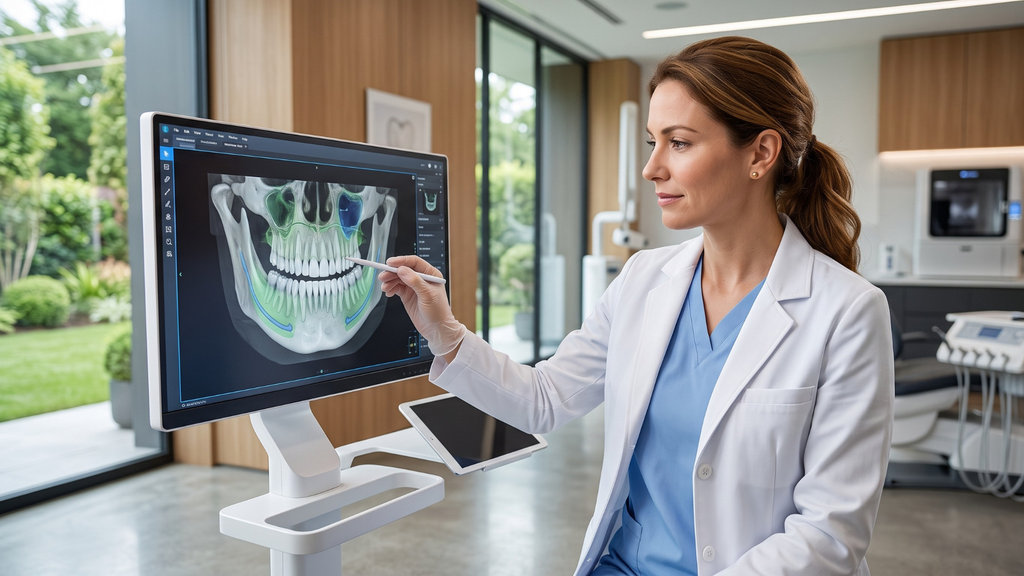
What an apicoectomy involves and what exactly is removed
The dentist lifts the gum and exposes the bone over the root. A window is opened in it, the inflamed tissue is removed and the root tip itself is cut off. About three millimetres goes. That is the figure given by the 2025 expert consensus, that is an agreed position of the Chinese dental society, and not the result of a single study. There was no European or American equivalent at the time. The reason is anatomical: the last three millimetres hold most of the lateral canals and ramifications, which cannot be cleaned from the crown side. Cutting off the tip does not in itself seal anything. The canal is left open from below, so it has to be prepared and filled from that side. This stage is called root-end preparation and root-end filling. The consensus recommends a preparation at least three millimetres deep, running along the canal and carried out with ultrasonics.
It happens that the lesion removed turns out to be a cyst, and what settles that is histopathological examination.
This is exactly what separates contemporary endodontic microsurgery from the older procedure at the root tip. The difference is made by high magnification, by a canal prepared from the root end with an ultrasonic tip, and by a biocompatible sealing material.
The name root resection can mislead, because it suggests removal of the whole root. That is a different procedure, carried out in multi-rooted teeth. Here it is the tip of the root that goes, and the tooth stays whole.
When an apicoectomy takes the place of retreatment
An apicoectomy is considered when access to the canal from above is closed off or too costly:
- the canal has become obliterated and cannot be negotiated;
- the canal is blocked by a post whose removal risks fracturing the root;
- filling material has been pushed beyond the root tip and the symptoms persist;
- the tooth carries a well-sealed crown and the lesion ends at the root tip itself.
Outside these situations, the first choice after failure remains root canal retreatment, because it removes the cause where the cause most often is, namely inside the canal.
The order is not symmetrical, though. Treatment from the crown side does not close off the route to a later procedure, whereas after the root tip has been cut off and the canal sealed from below, the reverse route can already be impassable. Nobody has measured how large that difference is; it follows from what is left of the root after the procedure.
Which route is more effective? The answer today is that it is not known. This is not for want of studies, but because they point in two directions. After the surgical procedure the lesion at the root tip appeared to heal more often, whereas the tooth stayed in place longer after nonsurgical treatment. A systematic review brought together five studies covering 529 teeth in all, followed from six months to just under nine years; it is described more fully in the text on root canal retreatment. The authors did not combine the results into a single figure: the studies were too different, and in four of the five the way they were conducted may have distorted the result. Neither method proved clearly better.
An analysis of insurance claims covering 1,021 teeth treated by endodontists reverses the direction of the figures, but it does not settle the matter either. After six years, 88 teeth in 100 were still in place after root-end surgery and 85 in 100 after retreatment. The difference falls within the bounds of chance, and in the retreatment group a later apicoectomy was also counted as a failure.
A decision table: retreatment, apicoectomy, extraction
The table sets out typical situations, but it does not replace an examination.
| Situation at the tooth | Route usually considered first | Why |
|---|---|---|
| Canal accessible, filling short or not sealed | root canal retreatment | the cause lies inside the canal and can be removed without surgery |
| Canal inaccessible: obliteration, a post, a well-sealed crown | apicoectomy | access from above costs more tissue than access from the gum |
| Material pushed beyond the root tip, symptoms persisting | apicoectomy | the lesion and the material lie outside the canal |
| A lesion at the root plus a deep pocket at the same tooth | a joint decision with periodontal treatment | sealing the canal alone does not remove the second cause |
| Vertical root fracture | removal of the tooth | the crack cannot be sealed from any side |
| Too little healthy tooth wall for a sealed restoration | removal of the tooth | a treated tooth will not hold the restoration anyway |
The last two rows open a separate conversation: save the tooth or place an implant.
How the procedure goes, step by step
An apicoectomy is one of the dental surgery procedures and is carried out under local anaesthetic, during a single appointment. The tooth being treated and the two teeth beside it are anaesthetised.
The order of the steps is fixed, although the studies have not counted the minutes for any of them:
- the dentist lifts a full-thickness gum flap covering the tooth being treated and the two teeth adjoining it;
- opens a window in the bone over the root tip, if the lesion has not already destroyed it, and removes the diseased tissue;
- cuts off about three millimetres of the root tip;
- prepares the canal from the root end with an ultrasonic tip, to a depth of at least three millimetres;
- fills that preparation with a biocompatible material, a calcium silicate-based bioceramic cement;
- sutures the gum, and removes the sutures after five to seven days.
Cone-beam computed tomography entered the standard of planning in endodontic microsurgery together with the microscope and piezoelectric instruments. It shows where the root tip lies in relation to the maxillary sinus and the mandibular canal, how thick the bone over the root is, and how far the lesion extends, which is precisely what an ordinary radiograph does not show accurately.
In the practice of Modern Dental & Orthodontics (Klinika MDO) it is repeatedly confirmed that patients ask about the brand of the material, as though the outcome depended on it. Studies point to something else. What counts is whether the root-end filling is sealed and deep enough. In a cohort of 148 out of 204 teeth, assessed with tomography from one to four years after the procedure, a correctly performed root-end filling was associated with a markedly greater chance of success. The figure given in that paper, more than fivefold, was calculated and compares the odds in the two groups; it does not say how many times more often the procedure succeeds.
Healing and aftercare: a timeline
An apicoectomy is over in the surgery on the same day, but its outcome is settled over the years that follow. Here is what happens when.
Day one. Pain reaches its peak. In a study covering 173 patients from a single practice, severe pain was reported by 17 in 100 people on the first day; over the following days the mean level of pain fell.
Days one to three. Swelling of the cheek and lip is at its greatest. Cooling from the outside with breaks through the first day, and sleeping with the head raised, are the standard recommendations. If the swelling lasts longer, the consensus permits warm compresses. Do not rinse your mouth vigorously and do not pull the lip back to inspect the wound.
Getting back to your activities. Once the swelling stops increasing, office work may be possible; exactly when depends on how large the swelling is. Physical exertion, saunas and contact sport are out at least until the sutures come out, because they raise the risk of bleeding and of the wound opening. Nobody has measured in studies how much time the return takes; this is a post-operative recommendation, not the result of a measurement.
Day five to seven. The sutures come out. Until then a soft diet applies, chewing on the opposite side and brushing as instructed, usually leaving out the gum margin itself.
Month three, six, twelve and twenty-four. Follow-up appointments with a radiograph.
Year four. The final assessment of the outcome. The radiograph after a year settles it provisionally; the bone goes on remodelling, and only after four years does the consensus treat the outcome as closed.
If a communication with the maxillary sinus occurred during the procedure, the instructions are stricter: do not blow your nose forcefully, do not swim and do not fly without your dentist's agreement, and sleep with your head raised. An antibiotic is then given for five to seven days. Outside that situation, none of the sources behind this text provides for an antibiotic as a routine after the procedure; whether one is needed is settled by the course of the procedure and by the patient's state of health.
How long the remodelling of the bone after an apicoectomy itself takes has not been measured: there is no study that would give that figure in months. The schedule of follow-up appointments therefore rests on practice, not on a measurement of the rate of healing.
Prognosis and possible complications
What an apicoectomy will give you is influenced by three factors that recur in the studies regardless of the centre. The first is periodontal involvement. In the cohort of 148 teeth assessed with tomography from one to four years after the procedure, the risk of failure where the lesion joined the focus at the root tip to periodontal disease was, on the same calculation as above, many times higher than where the lesion was confined to the root tip. The second factor is the state of the bone around the root. A pre-operative lesion with complete loss of the bone plate on the buccal side is listed by a review of prognostic factors among those reported most consistently. The third is the type of tooth: in the Swedish registry the odds of a mandibular molar being extracted within a decade were about two and a half times greater than for a maxillary incisor or canine. None of the three depends on how deftly the surgeon works, and all three are visible before the decision is taken.
The range given in the literature is itself wide. Survival of the tooth after surgery at the root tip falls in the studies between 48 and 93 teeth in 100, depending on how the patients were selected and on how long they were followed. A lesion unhealed on the radiograph and a tooth extracted are, moreover, two different outcomes: the first is counted by studies of healing, the second by registries that measure extraction alone. So the question to put to your dentist is this: does the figure you have quoted speak about the radiograph, or about the tooth?
To this is added the factor most often left out, which is time. A Spanish paper compared two groups of teeth from a single centre: with follow-up from one to four years, healing was found in 86.9% of 198 teeth, and with follow-up from five to nine years in 67.2% of 134. When teeth with a fractured root were excluded, the difference narrowed, but it did not disappear.
The consensus lists the complications by name: infection of the wound, damage to the root of the neighbouring tooth, perforation into the maxillary sinus, and disturbances of sensation, most often in the territory of the mental nerve. It does not state how often they occur. Nor does it answer the question that matters most to a patient faced with disturbed sensation: do they pass, or do they stay for good.
What is known, on the other hand, is how to tell that something is departing from the typical course: swelling that increases after the third day instead of subsiding, pain more severe than on the first day, fever, purulent discharge from the wound, or numbness of the lip persisting after the anaesthetic has worn off. Each of these is a reason to contact the dentist in charge of your treatment, not to wait for the appointment already booked.
Frequently asked questions
Does an apicoectomy hurt?
The procedure itself is carried out under local anaesthetic, so during it there is usually no pain. The discomfort comes afterwards and peaks on the first day: in a study covering 173 patients, severe pain was reported at that point by 17 in 100 people. Over the following days the pain eases; oral painkillers are used against the discomfort.
How long does the procedure itself take?
A single appointment under local anaesthetic, from the anaesthetic to the placing of the sutures. How long it will take depends on whether the tooth is a front tooth or a molar, on how many roots it has and on how thick the bone over the root tip is. A mandibular molar is technically the hardest. None of the studies this text rests on gives a typical time in minutes.
Will a visible scar be left after the procedure?
Usually no visible mark is left, but this depends on where the incision is placed. In front teeth the consensus recommends running it below the gum margin or at the base of the papilla, precisely in order to avoid the gum receding. Good healing of the gum is associated with better healing of the bone, so the appearance of the gum does say something about the outcome.
How long does the bone take to heal after an apicoectomy?
Longer than it takes to get back to normal functioning. The soft tissues heal in a few days, the bone takes considerably longer. Exactly how many months is not known: the studies do not give that figure. The consensus provides for follow-up appointments at three, six, twelve and twenty-four months, a provisional assessment of the outcome after a year, and a final one only after four years.
What are the chances that the procedure will succeed?
In observations running from one to four years, healing is found in close to nine teeth out of ten, and pooled across the whole literature in seven to eight. After five to nine years, one Spanish cohort gives 67.2%, and a Korean cohort followed for more than a decade gives 63.4% successes with 80.5% of teeth still present; that last figure is calculated on the part of the cohort that came back for follow-up.
Will a tooth after an apicoectomy hold a crown or a bridge?
Most often yes, because the crown and the post usually stay in place. The limit is set by a proportion: if, after the root tip has been cut off, the root in the bone would be shorter than the part of the tooth above the bone, the consensus treats that as a contraindication. A tooth supporting a bridge is additionally assessed for the load it carries.
What if the apicoectomy does not work?
A lesion unhealed on the radiograph after a year does not yet mean the tooth will be extracted: the consensus provides for a final assessment only after four years, because the lesion may still be shrinking during that time. If it is growing, or if the symptoms return, the same three routes are considered as before the procedure, extraction included. Whether a procedure already carried out makes a later implant harder is not settled by the studies in this set.
Can an apicoectomy be done at any tooth?
Not at any tooth. The obstacles include difficult access to the root tip, proximity to important anatomical structures, an acute phase of inflammation, poor oral hygiene, weak periodontal support, a vertical root fracture, and resorption of the root from the outside, that is loss of root tissue. The consensus requires general health to be assessed separately: chronic conditions and medicines, anticoagulants among them, have to be disclosed before the procedure. Blood clotting, among other things, is checked before surgery.
What to put in your calendar as you leave the surgery
The procedure takes one appointment. Assessing its outcome takes four years. What goes into the calendar, then, is the four follow-up appointments with a radiograph that the consensus provides for, at three, six, twelve and twenty-four months, and after them the most distant date of all, year four. Follow-up radiographs are part of the treatment, not a formality. Without them there is no knowing whether the lesion is receding.
Between the appointments, it is the course of healing that decides. Swelling that increases after the third day, fever, or numbness of the lip persisting after the anaesthetic has worn off are a reason to contact the practice, not to wait for the date already booked.
Read more:
- Dental Surgery in Warsaw Wola
- Root canal retreatment after failed treatment — when it makes sense and when it does not
- Dental cyst — what it is and why it should not be ignored
- Bone regeneration around teeth (GTR/GBR) — when a tooth can be saved instead of extracted
- Save the tooth or place an implant — how to make a decision you will not regret
Content and liability disclaimer
This article is informational and educational in nature and does not constitute medical advice, a diagnosis or a therapeutic recommendation — it does not replace a consultation with a specialist. If you are experiencing symptoms, have doubts or are facing a decision about treatment, consult a dentist. The methods described and the data cited are general in nature. The results of scientific studies relate to populations, not to an individual patient. The course and outcome of therapy depend on the individual clinical situation and may vary between patients. No information contained in this article constitutes a guarantee of result. The content has been prepared with due care, based on publicly available medical knowledge and the scientific publications indicated in the Sources section. We do not advise taking or refraining from any health-related action solely on the basis of the content of this article, without prior consultation with a doctor.
Sources
Source 1
Links https://doi.org/10.1038/s41368-024-00334-8 │ https://pubmed.ncbi.nlm.nih.gov/39743567/
Description Wang H, Xu X, Bian Z, Liang J, Chen Z, Hou B, Qiu L, Chen W, Wei X, Hu K i wsp. „Expert consensus on apical microsurgery.” International Journal of Oral Science. 2025;17(1):2.
Source 2
Links https://doi.org/10.1111/iej.13896 │ https://pubmed.ncbi.nlm.nih.gov/36710526/
Description Ng YL, Gulabivala K. „Factors that influence the outcomes of surgical endodontic treatment.” International Endodontic Journal. 2023;56(Suppl 2):116-139.
Source 3
Links https://doi.org/10.1111/iej.13783 │ https://pubmed.ncbi.nlm.nih.gov/35670053/
Description Setzer FC, Kratchman SI. „Present status and future directions: Surgical endodontics.” International Endodontic Journal. 2022;55(Suppl 4):1020-1058.
Source 4
Links https://doi.org/10.1111/iej.13793 │ https://pubmed.ncbi.nlm.nih.gov/35762859/
Description Bucchi C, Rosen E, Taschieri S. „Non-surgical root canal treatment and retreatment versus apical surgery in treating apical periodontitis: A systematic review.” International Endodontic Journal. 2023;56(Suppl 3):475-486.
Source 5
Links https://doi.org/10.1016/j.joen.2021.04.024 │ https://pubmed.ncbi.nlm.nih.gov/33961913/
Description Haxhia E, Ibrahim M, Bhagavatula P. „Root-end Surgery or Nonsurgical Retreatment: Are There Differences in Long-term Outcome?” Journal of Endodontics. 2021;47(8):1272-1277.
Source 6
Links https://doi.org/10.1016/j.joen.2024.04.009 │ https://pubmed.ncbi.nlm.nih.gov/38642732/
Description Yoo YJ, Cho EB, Perinpanayagam H, Gu Y, Zhu Q, Noblett WC, Kum KY. „Endodontic Microsurgery Outcomes over 10 Years and Associated Prognostic Factors: A Retrospective Cohort Study.” Journal of Endodontics. 2024;50(7):934-943.
Source 7
Links https://doi.org/10.1016/j.joen.2022.03.001 │ https://pubmed.ncbi.nlm.nih.gov/35292312/
Description Pallarés-Serrano A, Glera-Suarez P, Tarazona-Alvarez B, Peñarrocha-Oltra D, Peñarrocha-Diago M, Peñarrocha-Diago M. „Healing of 295 Endodontic Microsurgery Cases After Long-Term (5-9 Years) Versus Middle-Term (1-4 Years) Follow-up.” Journal of Endodontics. 2022;48(6):714-721.
Source 8
Links https://doi.org/10.1111/iej.13952 │ https://pubmed.ncbi.nlm.nih.gov/37403305/
Description Mota de Almeida FJ, Dawson VS, Kvist T, Frisk F, Bjørndal L, Isberg PE, Fransson H. „Periradicular surgery: a longitudinal registry study of ten-year outcomes and factors predictive of post-surgical extraction.” International Endodontic Journal. 2023;56(10):1212-1221.
Source 9
Links https://doi.org/10.1016/j.joen.2023.08.011 │ https://pubmed.ncbi.nlm.nih.gov/37633517/
Description Zhang MM, Fang GF, Wang ZH, Liang YH. „Clinical Outcome and Predictors of Endodontic Microsurgery Using Cone-beam Computed Tomography: A Retrospective Cohort Study.” Journal of Endodontics. 2023;49(11):1464-1471.
Source 10
Links https://doi.org/10.1016/j.joen.2020.12.012 │ https://pubmed.ncbi.nlm.nih.gov/33359530/
Description Malagise CJ, Khalighinejad N, Patel YT, Jalali P, He J. „Severe Pain after Endodontic Surgery: An Analysis of Incidence and Risk Factors.” Journal of Endodontics. 2021;47(3):409-414.