
A dental cyst is a lesion that can develop within the bone of the maxilla or mandible over a long period without symptoms. It is filled with fluid or semi-fluid content and is lined by epithelium. As it grows it gradually destroys the surrounding bone and may threaten adjacent teeth, nerves and the maxillary sinus.
Many patients are surprised by the diagnosis, because a cyst can remain painless for a long time. In clinical practice it is frequently detected incidentally during a routine radiographic examination — a panoramic radiograph or a CBCT scan taken for another reason.
Early detection of a cyst makes it possible to limit the extent of surgery, minimises the risk of complications and increases the chances of preserving the affected tooth. This article explains how a cyst forms, what symptoms it causes, how it is diagnosed and what treatment options are available.
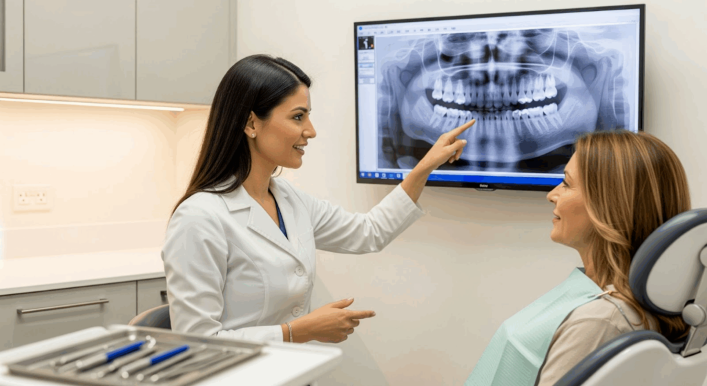
Symptoms of a dental cyst — how does the body signal the problem?
A dental cyst most commonly produces no symptoms — pain appears only when the cyst becomes infected or reaches a large size. The development of a cyst is usually slow, measured in months or years.
Local symptoms in the oral cavity
Most commonly we observe:
- a painless swelling of the gum or bone,
- a sensation of pressure in the maxilla or mandible,
- facial asymmetry when the cyst is large,
- mobility of the teeth adjacent to the lesion,
- displacement of the tooth crown,
- disturbance of tooth eruption (particularly canines and wisdom teeth).
Radiographic examination usually reveals a well-defined radiolucency in the bone, indicating bone loss.
Pain and inflammatory symptoms
Pain usually appears when the cyst becomes secondarily infected and an acute inflammatory process develops:
- throbbing pain in the region of the tooth or bone,
- facial or gingival swelling,
- redness of the mucosa,
- purulent discharge or a sinus tract,
- raised body temperature and general malaise.
In such cases prompt dental intervention is necessary.
Neurological symptoms
With larger cysts in the mandible, compression of the inferior alveolar nerve may occur. This manifests as:
- numbness of the lower lip,
- tingling of the chin,
- sensory disturbance in that area.
Symptoms of this kind require urgent imaging and a surgical or endodontic consultation.
The most common causes of dental cysts
The radicular cyst is the most common type — it accounts for 52–70% of all dental cysts and develops as a complication of untreated pulp necrosis. Other types are related to impacted teeth or developmental anomalies.
Radicular (inflammatory) cyst — a complication of untreated pulp necrosis
In clinical practice, a cyst most commonly develops as a consequence of chronic periapical inflammation. The process proceeds in stages:
- caries leads to infection of the pulp,
- pulp necrosis occurs and bacteria spread beyond the root,
- the chronic inflammatory process stimulates epithelial remnants in the periodontal ligament (Malassez rests),
- a cyst cavity filled with inflammatory fluid forms.
This type of lesion is frequently observed around teeth that underwent root canal treatment many years earlier.
Cyst associated with an impacted tooth
Cysts can develop around the crowns of teeth that have not erupted into the oral cavity (impacted teeth). This most commonly involves lower wisdom teeth and upper canines. A dentigerous (follicular) cyst accounts for 17–21% of dental cysts.
Developmental and keratinising cysts
Some cysts arise from disturbances in the development of odontogenic tissues. Of particular importance are keratinising cysts (keratocysts), which have a documented tendency to recur and require long-term follow-up after treatment.
Comparison of the most common types of dental cyst
The table below presents the key differences between the three most common types of dental cyst, facilitating an understanding of the diagnostics and treatment planning.
| Feature | Radicular cyst (inflammatory) | Dentigerous cyst (follicular) | Keratinising cyst (keratocyst) |
| Cause | Pulp necrosis, chronic inflammation | Impacted tooth eruption failure | Developmental anomaly of odontogenic tissue |
| Prevalence | 52–70% of all dental cysts | 17–21% of cysts | 5–17% of cysts |
| Typical location | Root apex of a non-vital tooth | Crown of an impacted tooth (wisdom teeth, canines) | Angle and body of the mandible |
| Radiographic / CBCT appearance | Round radiolucency at the apex | Radiolucency encompassing the tooth crown | Well-defined, multilocular |
| Treatment | Endodontics + cystectomy | Surgical removal together with the tooth | Cystectomy; requires follow-up (recurrences) |
| Tendency to recur | Low | Low | High — long-term follow-up required |
Why can a dental cyst be dangerous?
An untreated cyst gradually destroys bone and threatens adjacent anatomical structures. Complications include both local and systemic consequences.
Local consequences
An untreated cyst leads to:
- resorption of the maxillary or mandibular bone,
- displacement and loss of teeth,
- damage to sensory nerves of the face,
- abscess and sinus tract formation,
- perforation of the maxillary sinus.
In the case of very large lesions the bone may weaken, leading to a so-called pathological fracture.
Systemic significance
A chronic inflammatory process in the oral cavity places a burden on the body and may exacerbate the course of systemic diseases, including diabetes, cardiovascular disease and autoimmune conditions. This is an additional argument for not delaying treatment.
Diagnosis of a dental cyst: clinical and imaging investigations
The diagnosis of a dental cyst is based on a combination of clinical examination, radiographic imaging and histopathological analysis. Early detection frequently prevents extensive surgery.
Clinical examination
During the appointment the condition of the teeth, the periodontium (the tissues supporting the tooth in bone) and the mucosa is assessed. Tooth vitality and the presence of swelling or sinus tracts are checked.
Imaging diagnostics
- Periapical radiographs — suitable for small lesions.
- Panoramic radiograph — a screening panoramic examination.
- Cone-beam computed tomography (CBCT) — precise 3D visualisation of the cyst, assessment of bone loss and the relationship to nerves and sinuses.
CBCT is important in planning the surgical treatment of cysts. Studies demonstrate that CBCT detects periapical lesions that are invisible on conventional 2D radiographs.
Histopathological examination
The excised cyst is sent for microscopic examination, which confirms the type of lesion and excludes other pathologies, e.g. benign or malignant tumours.
Treatment of dental cysts — a modern therapeutic approach
The choice of treatment method depends on the type, size and location of the cyst. Small inflammatory lesions may resolve after root canal treatment, whereas larger cysts require surgical removal. Every treatment plan is individualised.
Endodontic treatment
Small inflammatory (radicular) cysts can be treated endodontically. The aim is to eliminate the source of infection and create conditions for bone regeneration.
Endodontic surgery
When conventional root canal treatment fails, root-end resection with simultaneous removal of the inflammatory lesion is performed. The procedure is planned on the basis of CBCT and uses microsurgical instruments.
Surgical removal of the cyst
Larger cysts require enucleation (cystectomy). The procedure is carried out under local anaesthesia and is planned on the basis of CBCT. In some cases augmentation of the resulting bone defect with a biomaterial is necessary.
Home remedies — as supportive treatment only
Analgesics, cold compresses and antiseptic rinses may alleviate inflammatory symptoms but do not eliminate the cyst. Delaying treatment increases the risk of complications.
Warning signs requiring an urgent dental appointment
Book an appointment immediately if you notice any of the following symptoms:
- rapidly increasing facial swelling,
- severe, throbbing pain,
- purulent discharge,
- numbness of the lip or chin,
- difficulty opening the mouth or swallowing.

Specialist consultation
The consultation includes thorough imaging diagnostics, an assessment of whether the tooth can be preserved, planning of the surgical or endodontic procedure, and a discussion of the treatment options and prognosis.
Recovery after dental cyst removal — practical advice
Healing after cyst removal proceeds in stages and requires adherence to post-operative instructions that reduce the risk of complications and support tissue regeneration.
During the first 2–3 days after the procedure, swelling and moderate pain are at their most intense. Cold compresses (20 minutes per hour) and analgesics prescribed by the clinician are helpful during this period.
For the first week a liquid or semi-liquid diet is recommended, avoiding hot foods and drinks. During oral hygiene the surgical area should be bypassed; gentle rinsing with chlorhexidine is usually recommended from the second day.
Soft-tissue healing usually takes 2–4 weeks. Bone regeneration after cyst removal may take from several months to over a year, depending on the size of the defect. The clinician monitors progress with follow-up radiographic examinations.
The most frequently asked questions about dental cysts
Does a dental cyst hurt?
Most commonly it causes no pain for a long time. Symptoms appear upon secondary infection or compression of nerve structures. The absence of pain does not mean that the cyst is not growing — regular radiographic check-ups are essential.
Can a dental cyst resolve on its own?
No. A cyst is a closed structure lined by epithelium that maintains its volume. Without treatment the lesion will gradually enlarge. Spontaneous disappearance of a true cyst has not been documented in the scientific literature.
How quickly does a dental cyst grow?
The growth rate depends on the type of cyst. Inflammatory cysts develop slowly, over several years, whereas developmental or keratinising cysts can grow faster — sometimes becoming clinically apparent within months.
Is a dental cyst dangerous?
Yes — it can lead to bone loss, tooth loss, nerve damage and sinus tract formation. A long-standing inflammatory process also places a burden on the body. Early detection and treatment minimise these risks.
Can a cyst be seen on a dental radiograph?
Small cysts are often detected on periapical radiographs, larger ones on a panoramic view. An important diagnostic tool is CBCT, which provides a three-dimensional image of the cyst and enables precise surgical planning.
What does dental cyst removal look like?
The procedure is performed under local anaesthesia and involves surgical enucleation (cystectomy) of the lesion from the bone. If necessary, root-end resection of the affected tooth or bone augmentation is carried out simultaneously.
Can the tooth be saved after cyst removal?
In many cases, yes, provided that sufficient tooth structure and bone support are preserved. Root canal treatment or root-end resection is often necessary, combined with long-term radiographic follow-up.
How long does bone healing take after cyst removal?
Soft-tissue healing usually takes 2–4 weeks. Bone regeneration is a prolonged process — from several months to over a year, depending on the size of the defect. The clinician monitors progress with follow-up imaging.
Can a dental cyst recur?
Some cysts, particularly keratinising cysts (keratocysts), have a documented tendency to recur. For this reason, long-term radiographic follow-up — usually every 6–12 months for the first 5 years — is recommended after treatment.
Summary
A dental cyst is a lesion within bone that can develop asymptomatically but in the long term leads to local complications and affects overall health. The key to successful treatment is early diagnosis (radiography, CBCT) and prompt intervention (endodontic or surgical).
At Modern Dental & Orthodontics we combine modern imaging diagnostics (radiography, CBCT) with the experience of a team of endodontists, oral surgeons and prosthodontists. Every case is assessed individually — so that the patient receives a treatment plan tailored precisely to their clinical situation.
Read more on the Modern Dental & Orthodontics website:
Dental Surgery Warsaw Wola | Make an appointment
Root canal treatment Warsaw Wola | Endodontics | MDO Clinic
Root canal treatment step by step — MDO Blog
Endodontics explained — MDO Blog
Dental radiography, panoramic and 3D CBCT — MDO Blog
Comprehensive dental check-up — MDO Blog
Scientific sources
Source 1
Link: https://pubmed.ncbi.nlm.nih.gov/39370695
Bibliographic description: Rees V, Klare M, Samaniego V, Leiva F, Jara R, Martínez Rondanelli B, González Providell S. "Epidemiological Features of Odontogenic Cysts in a Chilean Hospital: A 10-Year Retrospective Study." J Clin Med. 2024;13(19):5870.
Source 2
Link: https://pubmed.ncbi.nlm.nih.gov/31005378/
Bibliographic description: Mello FW, Melo G, Kammer PV, Speight PM, Rivero ERC. "Prevalence of odontogenic cysts and tumors associated with impacted third molars: A systematic review and meta-analysis." J Craniomaxillofac Surg. 2019;47(6):996-1002.
Source 3
Bibliographic description: Kriplani S, Sedani S, Patel A, Chandak M, Shirbhate U. "Surgical Management of Radicular Cyst With the Use of Advanced Platelet-Rich Fibrin and Bone Graft: A Case Report." Cureus. 2024;16(5):e61148.
Source 4
Link: https://pmc.ncbi.nlm.nih.gov/articles/PMC7455399/
Bibliographic description: Nik Abdul Ghani NR, Abdul Hamid NF, Karobari MI. „‘Tunnel’ radicular cyst and its management with root canal treatment and periapical surgery: A case report”. Clin Case Rep. 2020;8:1387–1391.
Information on content and responsibility
This article is intended solely for informational and educational purposes and does not constitute medical advice, a diagnosis or a treatment recommendation. It does not replace a consultation with a dentist or other qualified specialist. Despite every effort to ensure accuracy, the authors accept no liability for decisions made by readers on the basis of the information contained herein.