Introduction: a panoramic report that the parent was not expecting
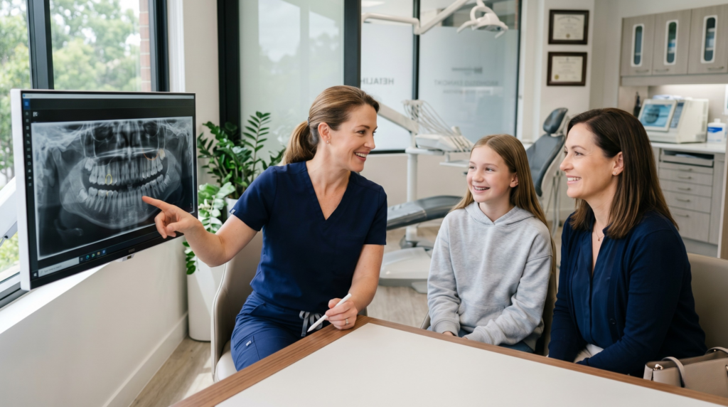
A thirteen-year-old patient is referred for a panoramic radiograph during the transition from primary to permanent teeth. A few days later the radiological report arrives, containing a phrase that sounds unfamiliar and alarming to the parent: "upper canines impacted in the palate, in close proximity to the roots of the lateral incisors". The question that we hear regularly in the clinic follows: can this still be reversed, will surgery be needed, and will the child have their own healthy teeth?
In most cases the answer to these questions is positive. Guiding an impacted canine into the dental arch (orthodontic extrusion, i.e. the controlled introduction of the tooth into the correct position) is a classic team-based treatment: the orthodontist designs the biomechanical plan, the oral and maxillofacial surgeon exposes the crown of the tooth, and the orthodontist gradually introduces the tooth into the arch over a period of twelve to twenty-four or so months. This article explains step by step when treatment is worthwhile, when extraction of the impacted tooth and future replacement with an implant is the more sensible decision, and what questions are worth asking before the first decision is made.
What is an impacted tooth — a clinical definition
An impacted tooth is a permanent tooth that has not erupted into the oral cavity within the expected time and no longer has the biological potential to do so spontaneously. The teeth most commonly impacted are third molars (wisdom teeth) and upper canines. The prevalence of upper canine impaction in the European population is estimated at 1–3 per cent. Management involves 3D diagnostics (CBCT), combined orthodontic-surgical planning and multistage team-based treatment.
Why canines are special and require separate attention
The upper canines play a key role in the dental arch: they are the longest teeth, the most firmly anchored in bone, they determine the width of the smile line and they govern correct masticatory mechanics (i.e. the way the upper and lower teeth meet during biting). This is why loss or incorrect positioning of a canine affects the entire aesthetic and functional outcome of orthodontic treatment.
The most common site of impaction is the palate — the canine lies deep within the palatal bone, usually at an oblique angle. The buccal side is a less frequent location. The mechanism of impaction is not fully understood, but the factors implicated include: an abnormal eruption path, lack of space in the arch, a mechanical obstacle (most commonly the root of the lateral incisor), genetic factors, and skeletal features related to the anatomy of the maxilla — as confirmed by the most recent meta-analysis in the European Journal of Orthodontics (Gudelevičiūtė et al., 2023).
An important point for parents: in most cases impaction is painless and produces no symptoms — it is discovered "incidentally" during a routine panoramic radiograph in a child aged 11–13. This is the moment at which time begins to work either in favour of or against the prognosis.
Why one cannot simply wait
This is one of the most frequently repeated questions in the clinic. The answer has two layers.
Firstly — a canine that has not erupted by the age of 14–15 will most likely never erupt spontaneously. The older the patient, the higher the risk of ankylosis, i.e. fusion of the tooth with the surrounding bone. Ankylosis dramatically reduces the chances of successful orthodontic extrusion into the arch.
Secondly, and more importantly — an impacted canine can cause resorption (progressive destruction of tissue) of the roots of adjacent teeth, most commonly the lateral incisors. A current meta-analysis published in Children (Mitsea et al., 2022) indicates that root resorption of adjacent teeth is detected significantly more often on CBCT than on conventional panoramic radiographs (OPG) — meaning that many cases remain undiagnosed when diagnostics are limited to two-dimensional imaging. Resorption is painless but progressive. This is the clinical rationale for why 3D diagnostics are not an "add-on" at our clinic but a standard of care whenever canine impaction is suspected.
Diagnostics: panoramic radiograph vs CBCT — a difference that changes the treatment plan
A conventional panoramic radiograph (OPG) is the first investigation that usually reveals the impaction. It shows the presence and general position of the tooth, but it is two-dimensional: it does not reliably answer the questions of how deep the canine lies, whether it is palatal or buccal, or whether resorption of the roots of adjacent teeth is present. A high-quality treatment plan requires answers to each of these questions.
These questions are resolved by cone-beam computed tomography (CBCT) — a three-dimensional radiological examination with a low radiation dose that allows the tooth to be viewed from every side, distances to be measured in millimetres and the risk of iatrogenic (procedure-related) damage to adjacent teeth to be assessed. From our orthodontic-surgical practice at Modern Dental & Orthodontics: CBCT before planning impacted canine extrusion is not a luxury but a minimum of due clinical diligence — a position supported by current systematic reviews (including Yi et al., BMC Oral Health 2026).
What exactly does the orthodontist see on CBCT?
- The position of the impacted canine in three dimensions — depth, direction of displacement (buccal or palatal), angle of inclination of the crown and root.
- The condition of the roots of adjacent teeth — presence and degree of resorption, location of any damage.
- The condition of the hard tissues surrounding the tooth, including the thickness of the buccal plate.
- The presence of additional structures (supernumerary teeth, cysts) — because tooth impaction is sometimes secondary to other abnormalities.
Three prognostic scenarios and the corresponding treatment plans
After CBCT analysis the treatment plan takes shape according to three main scenarios. This classification has practical significance: it allows the prognosis to be honestly defined and alternatives to be presented to the patient before a decision is made to embark on a multi-year process.
Scenario 1: Favourable impaction — full orthodontic-surgical treatment
The canine lies at a moderate depth, in a position with a good prognosis, there is no resorption of adjacent teeth, and the patient is aged 11–14. This is the classic indication for team-based treatment: the orthodontist fits fixed braces, prepares space in the arch, and then the oral and maxillofacial surgeon exposes the crown of the impacted canine and bonds an orthodontic attachment (a small metal button with a chain, known clinically as a button) to it. The orthodontist, over a period of several months, gradually introduces the canine to its correct position using controlled forces. Duration of the full process: 18–30 months. Success rate in selected groups: above 90%.
Scenario 2: Borderline impaction — treatment with TADs or a compromise approach
The canine lies deeper, but a prognosis still exists. Conventional biomechanics may be insufficient — in such cases orthodontic mini-screws (TADs) are used as absolute anchorage points (we discuss the details in our article on orthodontic mini-screws). Treatment is longer and the risk of complications (root resorption, ankylosis) is higher. The patient must be informed of both the realistic chances of success and the alternative scenario.
Scenario 3: Unfavourable impaction — extraction as a sensible alternative
The canine lies very deep, is strongly fused with the bone (signs of ankylosis visible on CBCT), the patient is an adult or an older adolescent (beyond the age of 16–17), and/or there is advanced resorption of adjacent roots. In such situations, attempting extrusion carries a high risk of failure and potential damage to the neighbouring teeth. Extraction of the impacted canine followed by prosthetic replacement — most commonly with a dental implant after growth is complete — may be the more responsible plan. This is a difficult conversation, but in the practice of the Modern Dental & Orthodontics team honesty about the prognosis forms the foundation of the therapeutic relationship.
The procedure: surgery + orthodontics step by step
Let us illustrate the full process using the favourable scenario — a 13-year-old patient, canine in a position with a good prognosis, team decision for full collaborative treatment. The stages are as follows.
Stage 1: Orthodontic preparation (3–9 months)
The orthodontist fits fixed braces on the remaining teeth. The appliance aligns the arch and opens space for the canine if necessary. This stage is indispensable — without adequate space in the arch the canine has nowhere to go, even after surgical exposure.
Stage 2: Surgical exposure of the canine (1 appointment, 30–60 minutes)
The oral and maxillofacial surgeon, under local anaesthesia, makes a gentle incision in the mucosa, reflects a soft-tissue flap, exposes the crown of the impacted canine and bonds an orthodontic button with a chain or an elastic ligature to its surface. The wound is then either closed (closed technique — the chain exits through a small opening in the suture) or left open (open technique). The choice of technique depends on the position of the canine.
Stage 3: Active extrusion of the canine (12–24 months)
At each follow-up appointment (usually every 4–6 weeks) the orthodontist applies a gentle elastic force through the chain, gradually guiding the canine towards its correct position in the arch. The process is slow and deliberate — excessively rapid forces damage the root and surrounding tissues. The patient sees the canine gradually emerging through the gum — a visually dramatic but biologically completely controlled process.
Stage 4: Final alignment and aesthetics (3–6 months)
Once the canine has reached the surface, the orthodontist positions it correctly in the arch, corrects rotation, angulation and occlusal contacts. In the final phase the goal is not merely function but an aesthetic result in which the erupted canine blends harmoniously with the line of the adjacent teeth.
Stage 5: Retention
After appliance removal, long-term retention is necessary — we discuss this in detail in our article on retention after orthodontics. In patients who have undergone impacted canine extrusion, a bonded retainer is almost always recommended, because the tooth has a natural tendency to drift back towards its original deep position.
Risks and limitations of treatment
- Failed extrusion — the canine does not respond to orthodontic traction. The most common cause is unrecognised ankylosis. In such situations the team makes a shared decision with the patient: extraction + prosthetic replacement or leaving the tooth monitored in situ.
- Root resorption of the lateral incisor — may worsen during orthodontic movements. This is why a follow-up CBCT (usually after 6–9 months of active extrusion) is an integral part of the protocol. If resorption progresses, the plan is modified.
- Gingival-line aesthetics — the canine guided into the arch may have a slightly different gum line from its counterpart on the healthy side; in selected cases minor periodontal surgery may be needed after treatment to level the gingival contour.
- Time and cost — the full treatment plan usually lasts 18–30 months and requires coordinated work between the orthodontist and the surgeon; the patient should receive an indicative cost estimate before the start of treatment.
FAQ — the most frequently asked questions about impacted canine treatment
Does an impacted canine hurt?
In the vast majority of cases — no. Canine impaction is painless and is discovered incidentally during a routine panoramic radiograph in a child aged 11–13. Symptoms (pain, swelling, an abscess) may appear only if the impacted tooth has caused an infection or a cyst — but these are rare complications of untreated impaction, not typical symptoms.
How long does it take to guide a canine into the arch?
The full orthodontic-surgical treatment plan usually takes 18–30 months and comprises four stages: orthodontic preparation (1–6 months), surgical exposure (1 appointment), active extrusion (12–24 months) and final alignment (3–6 months). Duration depends on the depth of the impacted canine, the patient's age and cooperation with the treatment plan.
Is the surgical canine exposure procedure painful?
The procedure itself is performed under local anaesthesia and the patient does not experience pain during it. After the anaesthesia wears off (usually 2–3 hours) moderate discomfort, swelling and slight sensitivity of the surgical area appear — these usually respond well to standard analgesics (paracetamol, ibuprofen) and resolve within 5–7 days.
Is CBCT really necessary if we already have a panoramic radiograph?
Yes — from the standpoint of patient safety and the quality of the treatment plan. A panoramic radiograph is a two-dimensional examination and does not reliably assess either the depth of impaction, the relationship to adjacent roots, or the presence of root resorption. CBCT provides this information with millimetre precision and, according to current evidence-based guidelines, is a standard of care in the diagnosis of impacted canines.
What happens if an impacted canine is left untreated?
In brief: the chance of introducing the tooth into the arch is lost, and the risk of complications increases. Over time ankylosis progresses (fusion of the tooth with bone), which permanently eliminates the possibility of orthodontic extrusion. Root resorption of the lateral incisors may worsen, and in rare cases a dentigerous cyst may develop around the impacted tooth. Monitoring every 12 months (CBCT) is an acceptable strategy only where the canine lies in a safe position and the patient has been fully informed of the risks.
Is there an alternative to full team-based treatment?
Yes — in selected situations. In children aged 9–12 in whom the canine is still in the eruption phase, extraction of the primary canine and orthodontic space creation sometimes prove effective — the permanent canine, finding room, erupts spontaneously. This is known as interceptive treatment and is only possible when caught early (which is one of the reasons why the first visit to the orthodontist is recommended at age 7). In adults, autotransplantation of the canine is sometimes considered, but this is a specialist procedure with limited indications.
Can I play sport during treatment?
In most cases, yes, with two caveats. After the surgical canine exposure procedure we recommend a 7–10 day break from intense exertion, contact sports and swimming. Throughout fixed braces treatment a mouthguard is recommended for contact sports. The orthodontist may issue specific recommendations depending on the stage of treatment.
Does canine extrusion affect the appearance of the smile?
Yes — and this is one of the most important reasons why this treatment is undertaken at all. Canines structure the aesthetics of the smile and the biomechanics of the bite. A correctly guided canine visually and functionally completes the arch. In some patients a minor difference in the gum line between the erupted canine and its counterpart on the opposite side may remain — if this is aesthetically significant, it can be corrected with a minor periodontal procedure after treatment.
Do I need to wear a retainer after treatment?
Yes — and in patients who have undergone impacted canine extrusion we recommend a bonded retainer (a wire cemented to the lingual surface of the teeth). A tooth that was deeply impacted in bone for years has a natural tendency to drift back towards its original position. A bonded retainer minimises this risk. In the practice of the Modern Dental & Orthodontics team we additionally recommend an Essix or Hawley retainer worn at night in both arches.
Read more:
- Orthodontist Warsaw Wola 🡪 klinikamdo.pl/en/offer/orthodontist/
- Dental surgery at Klinika MDO → klinikamdo.pl/en/offer/dental-surgery/
- Orthognathic surgery — when braces alone are not enough 🡪 https://klinikamdo.pl/en/blog/orthognathic-surgery-when-needed/
- Child grinds teeth at night — when is it an orthodontic problem? 🡪 https://klinikamdo.pl/en/blog/child-teeth-grinding-bruxism-orthodontics/
- Child breathes through the mouth — when is it a matter for the orthodontist? 🡪 https://klinikamdo.pl/en/blog/child-mouth-breathing-orthodontics/
- Wisdom teeth — remove or keep? A comprehensive extraction guide 🡪 https://klinikamdo.pl/en/blog/wisdom-teeth-removal-guide/
Sources
Source 1
Links https://doi.org/10.1186/s12903-026-08072-5 │ https://pubmed.ncbi.nlm.nih.gov/41807950/ │ https://www.ncbi.nlm.nih.gov/pmc/articles/PMC13088651/
Description Yi W, Abdullah JY, Mat Ali UM, Haque ASMR. „Diagnostic, predictive, and therapeutic approaches for impacted canines: a systematic review and meta-analysis.” BMC Oral Health. 2026;26(1).
Source 2
Links https://doi.org/10.3390/children9071006 │ https://pubmed.ncbi.nlm.nih.gov/35883990/ │ https://www.ncbi.nlm.nih.gov/pmc/articles/PMC9323464/
Description Mitsea A, Palikaraki G, Karamesinis K, Vastardis H, Gizani S, Sifakakis I. „Evaluation of Lateral Incisor Resorption Caused by Impacted Maxillary Canines Based on CBCT: A Systematic Review and Meta-Analysis.” Children (Basel). 2022;9(7):1006.
Source 3
Links https://doi.org/10.1002/14651858.CD012851.pub2 │ https://pubmed.ncbi.nlm.nih.gov/34967448/ │ https://www.ncbi.nlm.nih.gov/pmc/articles/PMC8717471/
Description Benson PE, Atwal A, Bazargani F, Parkin N, Thind B. „Interventions for promoting the eruption of palatally displaced permanent canine teeth, without the need for surgical exposure, in children aged 9 to 14 years.” Cochrane Database Syst Rev. 2021;12(12):CD012851.
Source 4
Links https://doi.org/10.1111/prd.12618 │ https://pubmed.ncbi.nlm.nih.gov/39548814/ │ https://onlinelibrary.wiley.com/doi/10.1111/prd.12618
Description Qali M, Li C, Chung CH, Tanna N. „Periodontal and orthodontic management of impacted canines.” Periodontol 2000. 2024.
Source 5
Links https://doi.org/10.1016/j.joms.2023.08.163 │ https://pubmed.ncbi.nlm.nih.gov/37683693/ │ https://www.sciencedirect.com/science/article/pii/S0278239123010285
Description Lwin CT, Cooney M, Goh M, Tham D, Nowak S. „Factors Associated With Successful Surgical Exposure of Impacted Maxillary Canines.” J Oral Maxillofac Surg. 2024;82(1):93-101.
Source 6
Links https://doi.org/10.1093/ejo/cjad050 │ https://pubmed.ncbi.nlm.nih.gov/37552898/ │ https://www.ncbi.nlm.nih.gov/pmc/articles/PMC10687515/
Description Gudelevičiūtė I, Spaičytė N, Smailienė D. „Skeletal and dental maxillary morphological characteristics in patients with impacted canines: systematic review and meta-analysis.” Eur J Orthod. 2023;45(6):832-841.