
Where magnification decides and where it merely helps
- Root canal treatment under the microscope is the same procedure carried out with a different view of the field. The microscope does not treat the tooth; it changes what the dentist sees at the moment of making a decision
- It makes the greatest difference where the problem is invisible to the naked eye: when searching for an extra canal, when an instrument has fractured inside the tooth, and when there is a crack in the root.
- Magnification has its price: the stronger it is, the smaller the part of the tooth that is in focus at any one time, and the slower the work goes.
- None of the studies this article rests on compares a whole course of treatment carried out with a microscope against one carried out without it. What is known is only how magnification behaves at individual stages.
- Isolation, irrigation and the operator's practised hand decide the outcome as well. Equipment on its own settles nothing.
The microscope has become a mark of quality in a practice. A patient who hears that the dentist works under magnification usually reads it as a promise that this time it will work — especially when the tooth has been treated once already and that treatment did not bring the expected result. The belief has a real basis; it is simply narrower than it is usually understood to be.
Magnification changes neither the biology of the infection nor the anatomy of the root. It changes one thing: how much the dentist sees when deciding where to place an instrument. At three of the five stages that difference carries over to the prognosis of the tooth; at the remaining two it may amount to no more than operator convenience, because nobody has measured what it contributes there.
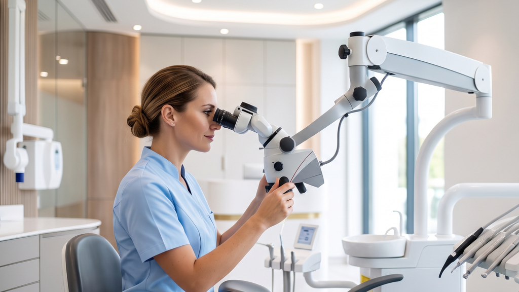
What the naked eye sees and what it does not
It all starts with one problem: the detail that settles the matter is smaller than what the eye can tell apart from the distance at which the dentist looks at the tooth.
The entrance to a root canal is usually a fraction of a millimetre across and lies a dozen or more millimetres deep inside the tooth, in a chamber shadowed by its own walls. The unaided eye tells apart detail on the order of tenths of a millimetre, and only in good light coming from the side, while the orifice of an extra canal, a fine line on the chamber floor or the edge of an old filling fall below that threshold.
The dental operating microscope, also called a dental microscope in the surgery, magnifies and illuminates at once. What strikes the patient is usually the magnification itself, but the light is often the more important half: it travels along the line of sight, so it falls on the very spot the dentist is looking at instead of on the wall beside it.
How much can be seen has been measured. A 2020 study compared three magnification settings of a dental operating microscope: the stronger the setting, the finer the detail that could be told apart, and the shallower the range that stayed in focus — about fifteen millimetres of depth at the weakest setting and about six at the strongest. At high magnification, then, the dentist sees a very thin slice of space very precisely. Eighteen people carried out a precision task on a phantom in the same study: twelve students and six dentists in specialist training, and under the microscope it took on average almost twice as long as with loupes. Something else, though, says the most. With every device, the dentists in training did better than the students. The equipment did not replace practised skill; it multiplied it.
We describe the basics of the procedure itself separately, in root canal treatment step by step.
The stages where magnification changes the most
The view becomes the bottleneck when the problem is small, hidden deep, and invisible without light delivered along the line of sight.
How to find every canal
The upper first molar, counted as the sixth tooth from the midline, most often has four canals. The fourth of them, MB2 for short, which is the second canal in the cheek-side front root, hides under a ledge of dentine on the floor of the chamber and is sometimes missed.
A missed canal has its price. A 2023 study based on cone-beam computed tomography compared two hundred and fifty-seven treated upper molars: those with the MB2 canal missed and those with it prepared. Where the canal had been missed, an inflammatory lesion at the root was seen distinctly more often: the calculated odds of it were about five and a half times higher. That does not mean such teeth develop the lesion five and a half times as often, because the measure used compares odds, not frequency itself. The size of the difference is uncertain in any case: the group was small enough that the calculation left room for a difference as modest as one and a half times and as large as nineteen times. The direction is certain. The magnitude is not.
Does magnification find that canal? In a 2023 laboratory study, on sixty-six extracted upper first molars with an MB2 canal confirmed on tomography, dentists looked for it with the naked eye, with loupes and under a microscope. No method found the canal in every tooth. Among operators under forty, magnification did not change the result; among older ones it changed it distinctly. This is a result from extracted teeth and from a small group; the authors themselves call their work preliminary, and magnification probably makes up for what the eye loses with age, namely the ability to focus at close range. That does not make the microscope redundant for a younger dentist, because in this one task the equipment alone simply was not enough. The microscope raises the chance of finding the canal, it does not give certainty, and the benefit is not spread evenly among operators.
An old root filling and a fractured instrument
Here the link between seeing and outcome is the most direct in the whole of endodontics. In a 2022 review of fractured instruments, the authors point out that the outcome is settled above all by whether the fragment can be seen. One that is visible can today be retrieved predictably; one that is not usually cannot. Magnification therefore moves some cases from one group to the other before anyone touches the tooth with an instrument. What that looks like in numbers is shown by a series published in 2023 covering thirty years of work by a single endodontist working under a microscope; out of one hundred and fifty-eight teeth, the fragment was retrieved from one hundred and thirty-one, which is roughly four cases in five. Those in which it succeeded were followed further: after five years fewer than eight in a hundred had failed, and every time the cause was a root fracture, not the instrument itself. Fragments lodged in the end portion of the root, near the apex, were harder. This is the result of one very experienced operator in his own practice, not an average across practices, and it has to be read that way.
A root fracture and a perforation
A vertical root fracture may be the one situation in which further root canal treatment makes no sense, because it usually ends with the tooth being removed. It is also the hardest of all to diagnose. The authors of a 2022 review of fractures put it plainly: tomography does not reliably show the crack itself, but it does show a characteristic pattern of bone loss around the root, and that raises the chance of a correct diagnosis and makes it possible to tell a fracture from an inflammatory lesion caused by something else. Clinical examination under magnification, with light along the line of sight, they regard as necessary. In this one place, magnification can be the tool that settles the fate of the tooth.
With a perforation, that is an accidental opening connecting the inside of the tooth with the tissues around the root, anatomy explains the advantage of magnification without difficulty, but measuring it is hard: there are no studies comparing the same repair with and without a microscope. In practical terms this means: "there are no studies" is not the same as "there is nothing to worry about", but neither is it the same as "nobody knows what to do". Repairing a perforation has its own rules; what has not been measured is only how much magnification adds to it.
Checking that the root filling is sealed
The last stage at which the question of the microscope arises is the end of treatment: whether the canal has been filled tightly and to the right length. That this matters is known. In a 2024 review of retreatment, the lesion at the root healed completely in fewer than eight teeth in ten, and the prognosis was better where the lesion before treatment was small or absent and where the canal was filled to the right length. A 2025 review adds the filling technique to that list. The contribution of magnification itself is measured by neither of these papers.
None of the ten studies this article rests on compares the outcome of treatment carried out under a microscope with the outcome of treatment carried out without one. All of them speak about single actions: finding a canal, retrieving a fragment, recognising a fracture. That is exactly where the division into stages comes from. Those ten papers cannot be assembled into a single figure saying how much a microscope raises the chance of saving a tooth — they simply answer a different question.
Five stages, and the state of the evidence for each:
| Stage of treatment | What the study showed | What it changes in the surgery |
|---|---|---|
| Searching for the MB2 canal | a missed canal is associated with higher odds of an inflammatory lesion; no method found it in every tooth | magnification raises the chance, it does not give certainty |
| Removing a fractured instrument | whether the procedure will go predictably is settled by whether the fragment can be seen | the microscope moves some cases from the "cannot be removed" group to "can be removed" |
| Vertical root fracture | tomography does not reliably show the crack; the authors regard examination under magnification as necessary | it can be the only way to tell a tooth to be treated from a tooth to be removed |
| Repairing a perforation | no studies comparing the procedure with and without magnification | the advantage follows from anatomy, not from measurement |
| Checking that the root filling is sealed | filling to the right length is associated with a better prognosis; the contribution of magnification is measured by none of these papers | length is settled by the radiograph, not by the dentist's eye |
When root canal treatment under a microscope gives no advantage
A single-rooted tooth with a wide, patent canal, treated for the first time for diagnosed irreversible pulpitis, rarely poses a problem that comes down to seeing; the bottleneck there is how tight the isolation is, how effective the irrigation is, and whether the tooth receives a sealed restoration in time.
Magnification improves none of those three things by itself.
Nor does the microscope look round a bend. It sees as far as light delivered along the line of sight reaches: to the canal orifice and to its straight portion, whereas the curved part of the root is viewed by nobody; that part of treatment rests on feel, on measuring the length and on the radiograph.
There are also times when magnification gets in the way. At a strong setting only a slice a few millimetres deep stays in focus, so the dentist refocuses every few moments. When opening the tooth, when placing the rubber dam and when rebuilding the crown, the dentist usually moves the microscope aside and works with loupes, because a wide field matters more then than resolution.
The claim "we use the microscope throughout the procedure" therefore says less than it seems to.
There is, finally, a limit that no magnification moves. The microscope does not change how bacteria behave inside the canal — we write about that in why root canal treatment is sometimes challenging.
The microscope is not everything: rubber dam, tomography and the dentist's hand
A microscope rarely stands in a surgery on its own. In a 2023 survey of more than five hundred endodontists from six countries, more than nine in ten used one; almost as many reported cone-beam tomography and an apex locator, and a rubber dam, the sheet of rubber isolating the tooth from saliva, was always used by nearly a hundred per cent. Those surveyed, however, were members of endodontic societies, a group better equipped than practices at large.
What practices call microscopic endodontics is in practice a whole protocol, with magnification only one part of it.
In a 2025 review gathering treatment outcomes assessed by tomography, the authors identified factors associated with success: the operator's experience, the number of appointments, and technical details of preparing and filling the canal — with experience standing there alongside the technical parameters, not below them.
There is one more thing in that paper that is rarely mentioned: the proportion of successes depends on how success is defined. Under the loose criteria, which accept a reduction in the lesion, a favourable result was recorded for roughly eight and a half teeth in ten; under the strict criteria, which require complete healing on tomography, for fewer than half. The same procedure, the same group of teeth, two different numbers. So when a practice quotes its own success rate, it is worth asking what exactly it counted as a success, because without that the figure alone says little about what a patient should expect for their own tooth.
In a 2025 position statement, the teachers of endodontology of the British Endodontic Society treated loupes as a minimum requirement as early as undergraduate level. That is an agreed expert opinion, not a study result, but it shows the direction: magnification is ceasing to be a distinguishing feature and becoming a starting point.
What the cost of root canal treatment under a microscope depends on
Quotations for the same tooth can differ several times over between practices, and it is almost never the microscope that does it; it is the number of canals, the history of the tooth and the extent of the restoration, which cannot be established before the tooth is opened. The question of how much root canal treatment under a microscope costs cannot be answered with a single figure: the cost of root canal treatment under a microscope depends on the tooth and on the practice, and a range given in advance would be misleading. Below are six things that shift that quotation.
- The number of canals and their anatomy. A molar with four canals is a different piece of work from an incisor with one.
- Whether the tooth has been treated before. Removing an old root filling, a post or a fractured instrument is a separate stage, sometimes longer than the treatment itself.
- The time set aside for the appointment. Working under magnification is slower, and that is a cost built into it.
- Diagnostics. Tomography is sometimes needed before treatment and is charged separately.
- The restoration afterwards. Its extent is decided once the tooth has been opened and it is clear how much healthy tissue is left.
- Breaking the quotation down into items. Ask for separate amounts for treating the canal, for the tomography and for the restoration. A quotation given over the telephone usually covers only the first of them.
In conversations with patients coming to Modern Dental & Orthodontics (Klinika MDO), one question returns: why two quotations for the same tooth differ so much. The answer nearly always lies in the diagnostics and in the extent of the restoration, not in the microscope. We describe the scope of the service on the page about root canal treatment.
What to ask a practice before treatment
- Will the procedure be carried out under rubber dam isolation? The answer "always" means more than "when possible".
- At which stages is the microscope used? A sensible answer points to stages rather than declaring use throughout.
- Will I receive photographs from the microscope taken during treatment? That is the only way to check the answer to the previous question.
- Is tomography needed, and who will report it?
- What happens if the canal turns out to be blocked? A practice with a fallback plan will set it out before treatment.
- Who will carry out the restoration, and how soon after treatment ends? A tooth without a sealed restoration loses what the treatment gave it.
Frequently asked questions
Does every tooth need treatment under a microscope?
No. With a wide, patent canal treated for the first time, the outcome is decided by isolation, irrigation and a sealed restoration, not by magnification. The microscope becomes necessary where the problem is invisible: when searching for an extra canal, in retreatment, and when a root fracture is suspected.
Is it worth paying extra for treatment under a microscope?
It depends on the tooth. With a tooth being treated for the first time, with one wide canal, the extra payment mainly buys comfort of work. In retreatment, with a fractured instrument and with a suspected root fracture, it buys the possibility of seeing a problem that cannot be seen without magnification. Before you ask about the amount, ask which of those situations your tooth belongs to.
Does root canal treatment under a microscope take longer?
Usually yes. In a phantom study, a precision task carried out under a microscope took on average almost twice as long as the same task with loupes. In the surgery this translates into a longer appointment or a larger number of them, depending on the tooth and on the treatment plan adopted.
Does treatment under a microscope hurt more?
Pain depends on the anaesthetic and on the state of the pulp, not on magnification. The microscope stands above you and does not touch your face; the dentist works further from the chair than usual. Working under magnification does, however, tend to take longer, so you have to hold out longer with your mouth open and the rubber dam in place. If you cope badly with long appointments, say so before treatment.
Does the microscope settle whether treatment succeeds?
No. What is associated with successful treatment is the operator's experience, the size of the lesion at the root before treatment, the length of the root filling and the way the canal was prepared. Root canal treatment under a microscope helps to do several of those things more precisely, but it replaces none of them. The proportion of successes itself depends on the measure adopted: under the loose criteria it is more than eight teeth in ten, under the strict criteria fewer than half.
How does a microscope differ from magnifying loupes?
In the range of magnification and in the light. Loupes give a fixed, modest magnification and illumination close to the line of sight. A microscope allows the magnification to be changed and delivers light exactly along that line, so it shows the inside of a narrow chamber. The price is a smaller depth over which the image stays in focus, and slower work.
Is treatment under a microscope reimbursed?
Guaranteed benefits cover root canal treatment within a defined scope and for selected groups of teeth, and the equipment of a surgery is not priced separately within them. In practice, treatment carried out under magnification usually takes place outside that scope. The scope of the guaranteed benefit at a particular practice has to be asked about directly before treatment begins.
What the microscope will not see for the dentist
Root canal treatment under a microscope is decisive in three places: when searching for a canal that cannot be seen, with a fractured instrument, and with a crack in the root. Beyond them, magnification is a convenience and a saving on error, not a condition of success. The question "do you have a microscope" is worse than the question "at which stages do you use it, and what do you do when it is not enough" — the first can be ticked off by buying equipment, the second requires the practice to describe how it actually works. The microscope shows what the dentist points it at, and not a millimetre more.
Read more:
- Root Canal Treatment in Warsaw
- Root canal treatment step by step: How long does it take, what does it look like and what to expect?
- Why is root canal treatment sometimes challenging?
- Endodontics explained. We answer patients' most difficult questions
- Pulpitis - reversible or irreversible. Why this distinction decides the fate of the tooth
Content and liability disclaimer
This article is informational and educational in nature and does not constitute medical advice, a diagnosis or a therapeutic recommendation — it does not replace a consultation with a specialist. If you are experiencing symptoms, have doubts or are facing a decision about treatment, consult a dentist. The methods described and the data cited are general in nature. The results of scientific studies relate to populations, not to an individual patient. The course and outcome of therapy depend on the individual clinical situation and may vary between patients. No information contained in this article constitutes a guarantee of result. The content has been prepared with due care, based on publicly available medical knowledge and the scientific publications indicated in the Sources section. We do not advise taking or refraining from any health-related action solely on the basis of the content of this article, without prior consultation with a doctor.
Sources
Source 1
Links https://doi.org/10.1016/j.joen.2020.01.013 │ https://pubmed.ncbi.nlm.nih.gov/32139266/
Description Al Shaikhly B, Harrel SK, Umorin M, Augsburger RA, Jalali P. „Comparison of a Dental Operating Microscope and High-resolution Videoscope for Endodontic Procedures.” Journal of Endodontics. 2020;46(5):688-693.
Source 2
Links https://doi.org/10.1111/aej.12729 │ https://pubmed.ncbi.nlm.nih.gov/36537343/
Description Olbertz J, Braguini AP, Vitali FC, Dos Santos JD, Martins HC, Régis JR, do Prado M, da Silveira Tiecher PF, Duque TM. „Influence of age and experience with magnification on locating the second mesiobuccal canal in maxillary first molars: A preliminary study.” Australian Endodontic Journal. 2023;49(Suppl 1):259-264.
Source 3
Links https://doi.org/10.1111/aej.12672 │ https://pubmed.ncbi.nlm.nih.gov/35908285/
Description Colakoglu G, Kaya Buyukbayram I, Elcin MA, Garip Berker Y, Ercalik Yalcinkaya S. „Association between second mesiobuccal canal and apical periodontitis in retrospective cone-beam computed tomographic images.” Australian Endodontic Journal. 2023;49(1):20-26.
Source 4
Links https://doi.org/10.1111/iej.13743 │ https://pubmed.ncbi.nlm.nih.gov/35377473/
Description Terauchi Y, Ali WT, Abielhassan MM. „Present status and future directions: Removal of fractured instruments.” International Endodontic Journal. 2022;55(Suppl 3):685-709.
Source 5
Links https://doi.org/10.1016/j.joen.2023.06.007 │ https://pubmed.ncbi.nlm.nih.gov/37422250/
Description Malentacca A, Zaccheo F, Rupe C, Lajolo C. „Endodontic Clinical Outcome after Separated Instrument Removal Using a Spinal Needle Technique: A Retrospective Study of Thirty Years of Clinical Experience.” Journal of Endodontics. 2023;49(8):980-989.
Source 6
Links https://doi.org/10.1016/j.joen.2024.01.013 │ https://pubmed.ncbi.nlm.nih.gov/38280514/
Description Sabeti M, Chung YJ, Aghamohammadi N, Khansari A, Pakzad R, Azarpazhooh A. „Outcome of Contemporary Nonsurgical Endodontic Retreatment: A Systematic Review of Randomized Controlled Trials and Cohort Studies.” Journal of Endodontics. 2024;50(4):414-433.
Source 7
Links https://doi.org/10.1111/iej.13737 │ https://pubmed.ncbi.nlm.nih.gov/35338655/
Description Patel S, Bhuva B, Bose R. „Present status and future directions: vertical root fractures in root filled teeth.” International Endodontic Journal. 2022;55(Suppl 3):804-826.
Source 8
Links https://doi.org/10.1016/j.joen.2025.03.004 │ https://pubmed.ncbi.nlm.nih.gov/40122230/
Description Brochado Martins JF, Georgiou AC, Nunes PD, de Vries R, Afreixo VMA, da Palma PJR, Shemesh H. „CBCT-Assessed Outcomes and Prognostic Factors of Primary Endodontic Treatment and Retreatment: A Systematic Review and Meta-Analysis.” Journal of Endodontics. 2025;51(6):687-706.
Source 9
Links https://doi.org/10.1111/iej.13982 │ https://pubmed.ncbi.nlm.nih.gov/37800848/
Description Cheung MC, Peters OA, Parashos P. „Global survey of endodontic practice and adoption of newer technologies.” International Endodontic Journal. 2023;56(12):1517-1533.
Source 10
Links https://doi.org/10.1111/iej.14153 │ https://pubmed.ncbi.nlm.nih.gov/39380470/
Description McLean W, Qualtrough AJE. „Teaching of magnification in the undergraduate curriculum: A position statement from the British Endodontic Society Teachers of Endodontology Group.” International Endodontic Journal. 2025;58(1):2-5.